4XYY

Hen Egg-White Lysozyme (HEWL) complexed with Zr(IV)-substituted Keggin-type polyoxometalate


Experimental Data Snapshot

  • Method: X-RAY DIFFRACTION
  • Resolution: 1.38 Å
  • R-Value Free: 0.184 
  • R-Value Work: 0.162 
  • R-Value Observed: 0.163 

Starting Model: experimental
View more details

wwPDB Validation   3D Report Full Report


Ligand Structure Quality Assessment 


This is version 1.3 of the entry. See complete history


Literature

Structural Characterization of the Complex between Hen Egg-White Lysozyme and Zr(IV) -Substituted Keggin Polyoxometalate as Artificial Protease.

Sap, A.De Zitter, E.Van Meervelt, L.Parac-Vogt, T.N.

(2015) Chemistry 21: 11692-11695

  • DOI: https://doi.org/10.1002/chem.201501998
  • Primary Citation of Related Structures:  
    4XYY

  • PubMed Abstract: 

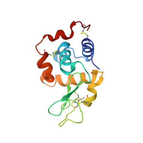
    Successful co-crystallization of a noncovalent complex between hen egg-white lysozyme (HEWL) and the monomeric Zr(IV) -substituted Keggin polyoxometalate (POM) (Zr1 K1), (Et2 NH2)3 [Zr(PW11 O39)] (1), has been achieved, and its single-crystal X-ray structure has been determined. The dimeric Zr(IV) -substituted Keggin-type polyoxometalate (Zr1 K2), (Et2 NH2)10 [Zr(PW11 O39 )2] (2), has been previously shown to exhibit remarkable selectivity towards HEWL hydrolysis. The reported X-ray structure shows that the hydrolytically active Zr(IV) -substituted Keggin POM exists as a monomeric species. Prior to hydrolysis, this monomer interacts with HEWL in the vicinity of the previously identified cleavage sites found at Trp28-Val29 and Asn44-Arg45, through water-mediated H-bonding and electrostatic interactions. Three binding sites are observed at the interface of the negatively charged Keggin POM and the positively charged regions of HEWL at: 1) Gly16, Tyr20, and Arg21; 2) Asn44, Arg45, and Asn46; and 3) Arg128.


  • Organizational Affiliation

    Department of Chemistry, KU Leuven, Celestijnenlaan 200F box 2404, 3001 Leuven, Heverlee (Belgium).


Macromolecules
Find similar proteins by:  (by identity cutoff)  |  3D Structure
Entity ID: 1
MoleculeChains Sequence LengthOrganismDetailsImage
Lysozyme C129Gallus gallusMutation(s): 0 
EC: 3.2.1.17
UniProt
Find proteins for P00698 (Gallus gallus)
Explore P00698 
Go to UniProtKB:  P00698
Entity Groups  
Sequence Clusters30% Identity50% Identity70% Identity90% Identity95% Identity100% Identity
UniProt GroupP00698
Sequence Annotations
Expand
  • Reference Sequence
Experimental Data & Validation

Experimental Data

  • Method: X-RAY DIFFRACTION
  • Resolution: 1.38 Å
  • R-Value Free: 0.184 
  • R-Value Work: 0.162 
  • R-Value Observed: 0.163 
  • Space Group: P 43 21 2
Unit Cell:
Length ( Å )Angle ( ˚ )
a = 77.232α = 90
b = 77.232β = 90
c = 37.961γ = 90
Software Package:
Software NamePurpose
PHENIXrefinement
PDB_EXTRACTdata extraction
XDSdata reduction
PHASERphasing
XDSdata scaling

Structure Validation

View Full Validation Report


Ligand Structure Quality Assessment 


Entry History 

Deposition Data

Revision History  (Full details and data files)

  • Version 1.0: 2015-07-29
    Type: Initial release
  • Version 1.1: 2015-08-12
    Changes: Database references
  • Version 1.2: 2018-12-12
    Changes: Data collection, Source and taxonomy, Structure summary
  • Version 1.3: 2024-01-10
    Changes: Data collection, Database references, Refinement description