4XXO | pdb_00004xxo

Crystal Structure of Human APOBEC3A


Experimental Data Snapshot

  • Method: X-RAY DIFFRACTION
  • Resolution: 2.84 Å
  • R-Value Free: 
    0.253 (Depositor), 0.257 (DCC) 
  • R-Value Work: 
    0.214 (Depositor), 0.221 (DCC) 
  • R-Value Observed: 
    0.217 (Depositor) 

Starting Model: experimental
View more details

wwPDB Validation 3D Report Full Report

Validation slider image for 4XXO

This is version 1.6 of the entry. See complete history

Literature

The ssDNA Mutator APOBEC3A Is Regulated by Cooperative Dimerization.

Bohn, M.F.Shandilya, S.M.Silvas, T.V.Nalivaika, E.A.Kouno, T.Kelch, B.A.Ryder, S.P.Kurt-Yilmaz, N.Somasundaran, M.Schiffer, C.A.

(2015) Structure 23: 903-911

  • DOI: https://doi.org/10.1016/j.str.2015.03.016
  • Primary Citation Related Structures: 
    4XXO

  • PubMed Abstract: 

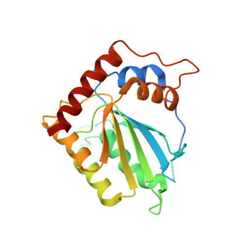
    Deaminase activity mediated by the human APOBEC3 family of proteins contributes to genomic instability and cancer. APOBEC3A is by far the most active in this family and can cause rapid cell death when overexpressed, but in general how the activity of APOBEC3s is regulated on a molecular level is unclear. In this study, the biochemical and structural basis of APOBEC3A substrate binding and specificity is elucidated. We find that specific binding of single-stranded DNA is regulated by the cooperative dimerization of APOBEC3A. The crystal structure elucidates this homodimer as a symmetric domain swap of the N-terminal residues. This dimer interface provides insights into how cooperative protein-protein interactions may affect function in the APOBEC3 enzymes and provides a potential scaffold for strategies aimed at reducing their mutation load.


  • Organizational Affiliation
    • Department of Biochemistry and Molecular Pharmacology, University of Massachusetts Medical School Worcester, 364 Plantation Street, MA 01605, USA.

Macromolecule Content 

  • Total Structure Weight: 47.13 kDa 
  • Atom Count: 3,091 
  • Modeled Residue Count: 373 
  • Deposited Residue Count: 406 
  • Unique protein chains: 1

Macromolecules

Find similar proteins by:|  3D Structure
Entity ID: 1
MoleculeChains  Sequence LengthOrganismDetailsImage
DNA dC->dU-editing enzyme APOBEC-3A
A, B
203Homo sapiensMutation(s): 2 
Gene Names: APOBEC3A
EC: 3.5.4 (PDB Primary Data), 3.5.4.38 (UniProt)
UniProt & NIH Common Fund Data Resources
Find proteins for P31941 (Homo sapiens)
Explore P31941 
Go to UniProtKB:  P31941
PHAROS:  P31941
GTEx:  ENSG00000128383 
Entity Groups
Sequence Clusters30% Identity50% Identity70% Identity90% Identity95% Identity100% Identity
UniProt GroupP31941
Sequence Annotations
Expand
Reference Sequence

Small Molecules

Ligands 2 Unique
IDChains Name / Formula / InChI Key2D Diagram3D Interactions
ZN

Query on ZN


Download:Ideal Coordinates CCD File
C [auth A]
D [auth A]
E [auth A]
F [auth A]
G [auth A]
C [auth A],
D [auth A],
E [auth A],
F [auth A],
G [auth A],
I [auth B],
J [auth B]
ZINC ION
Zn
PTFCDOFLOPIGGS-UHFFFAOYSA-N
CL

Query on CL


Download:Ideal Coordinates CCD File
H [auth A],
K [auth B]
CHLORIDE ION
Cl
VEXZGXHMUGYJMC-UHFFFAOYSA-M

Experimental Data & Validation

Experimental Data

  • Method: X-RAY DIFFRACTION
  • Resolution: 2.84 Å
  • R-Value Free:  0.253 (Depositor), 0.257 (DCC) 
  • R-Value Work:  0.214 (Depositor), 0.221 (DCC) 
  • R-Value Observed: 0.217 (Depositor) 
Space Group: P 65 2 2
Unit Cell:
Length ( Å )Angle ( ˚ )
a = 94.752α = 90
b = 94.752β = 90
c = 213.962γ = 120
Software Package:
Software NamePurpose
PHENIXrefinement
HKL-3000data reduction
HKL-3000data scaling
PHENIXphasing

Structure Validation

View Full Validation Report


Entry History 

& Funding Information

Deposition Data


Funding OrganizationLocationGrant Number
National Institutes of Health/National Institute of General Medical Sciences (NIH/NIGMS)United StatesGM091743

Revision History  (Full details and data files)

  • Version 1.0: 2015-04-29
    Type: Initial release
  • Version 1.1: 2015-05-13
    Changes: Database references
  • Version 1.2: 2015-05-20
    Changes: Database references
  • Version 1.3: 2017-09-27
    Changes: Author supporting evidence, Database references, Derived calculations, Source and taxonomy
  • Version 1.4: 2019-12-25
    Changes: Author supporting evidence
  • Version 1.5: 2023-09-27
    Changes: Data collection, Database references, Derived calculations, Refinement description
  • Version 1.6: 2024-11-06
    Changes: Structure summary