Exploring Multimodularity in Plant Cell Wall Deconstruction: STRUCTURAL AND FUNCTIONAL ANALYSIS OF Xyn10C CONTAINING THE CBM22-1-CBM22-2 TANDEM.
Sainz-Polo, M.A., Gonzalez, B., Menendez, M., Pastor, F.I., Sanz-Aparicio, J.(2015) J Biological Chem 290: 17116-17130
- PubMed: 26001782
- DOI: https://doi.org/10.1074/jbc.M115.659300
- Primary Citation of Related Structures:
4W8L, 4XUN, 4XUO, 4XUP, 4XUQ, 4XUR, 4XUT - PubMed Abstract:
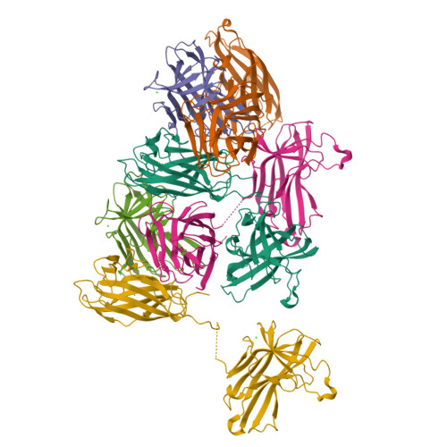
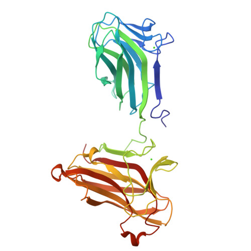
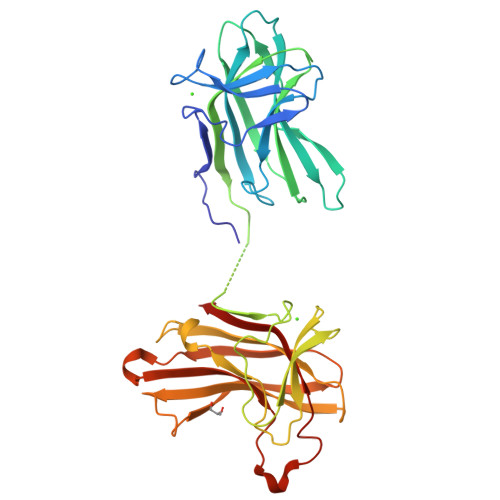
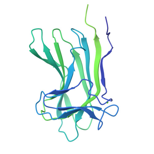
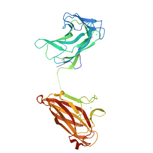
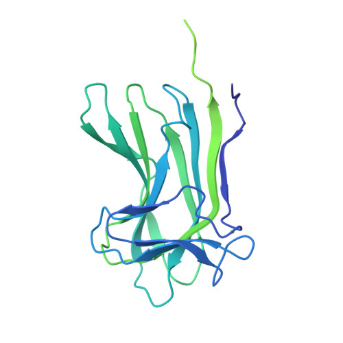
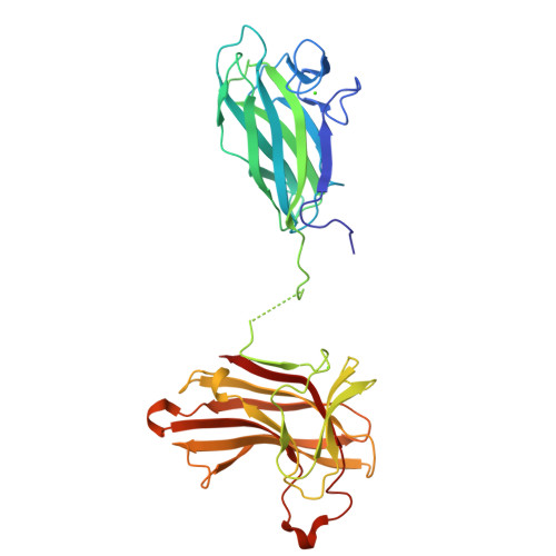
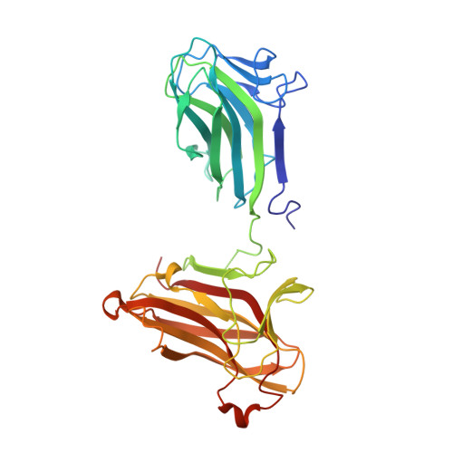
Elucidating the molecular mechanisms regulating multimodularity is a challenging task. Paenibacillus barcinonensis Xyn10C is a 120-kDa modular enzyme that presents the CBM22/GH10/CBM9 architecture found in a subset of large xylanases. We report here the three-dimensional structure of the Xyn10C N-terminal region, containing the xylan-binding CBM22-1-CBM22-2 tandem (Xyn10C-XBD), which represents the first solved crystal structure of two contiguous CBM22 modules. Xyn10C-XBD is folded into two separate CBM22 modules linked by a flexible segment that endows the tandem with extraordinary plasticity. Each isolated domain has been expressed and crystallized, and their binding abilities have been investigated. Both domains contain the R(W/Y)YYE motif required for xylan binding. However, crystallographic analysis of CBM22-2 complexes shows Trp-308 as an additional binding determinant. The long loop containing Trp-308 creates a platform that possibly contributes to the recognition of precise decorations at subsite S2. CBM22-2 may thus define a subset of xylan-binding CBM22 modules directed to particular regions of the polysaccharide. Affinity electrophoresis reveals that Xyn10C-XBD binds arabinoxylans more tightly, which is more apparent when CBM22-2 is tested against highly substituted xylan. The crystal structure of the catalytic domain, also reported, shows the capacity of the active site to accommodate xylan substitutions at almost all subsites. The structural differences found at both Xyn10C-XBD domains are consistent with the isothermal titration calorimetry experiments showing two sites with different affinities in the tandem. On the basis of the distinct characteristics of CBM22, a delivery strategy of Xyn10C mediated by Xyn10C-XBD is proposed.
Organizational Affiliation:
From the Departamentos de Cristalografía y Biología Estructural y.


