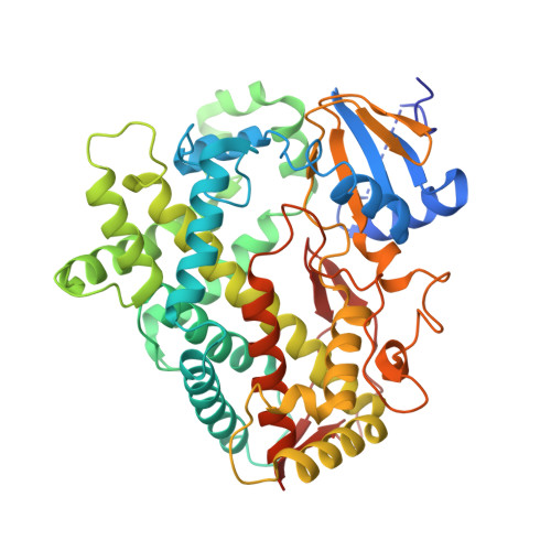
4XRZ

Human Cytochrome P450 2D6 BACE1 Inhibitor 6 Complex


Experimental Data Snapshot

  • Method: X-RAY DIFFRACTION
  • Resolution: 2.40 Å
  • R-Value Free: 0.254 
  • R-Value Work: 0.217 
  • R-Value Observed: 0.219 

Starting Model: experimental
View more details

wwPDB Validation   3D Report Full Report


Ligand Structure Quality Assessment 


This is version 1.3 of the entry. See complete history


Literature

Utilizing Structures of CYP2D6 and BACE1 Complexes To Reduce Risk of Drug-Drug Interactions with a Novel Series of Centrally Efficacious BACE1 Inhibitors.

Brodney, M.A.Beck, E.M.Butler, C.R.Barreiro, G.Johnson, E.F.Riddell, D.Parris, K.Nolan, C.E.Fan, Y.Atchison, K.Gonzales, C.Robshaw, A.E.Doran, S.D.Bundesmann, M.W.Buzon, L.Dutra, J.Henegar, K.LaChapelle, E.Hou, X.Rogers, B.N.Pandit, J.Lira, R.Martinez-Alsina, L.Mikochik, P.Murray, J.C.Ogilvie, K.Price, L.Sakya, S.M.Yu, A.Zhang, Y.O'Neill, B.T.

(2015) J Med Chem 58: 3223-3252

  • DOI: https://doi.org/10.1021/acs.jmedchem.5b00191
  • Primary Citation of Related Structures:  
    4XRY, 4XRZ, 4XXS

  • PubMed Abstract: 

    In recent years, the first generation of β-secretase (BACE1) inhibitors advanced into clinical development for the treatment of Alzheimer's disease (AD). However, the alignment of drug-like properties and selectivity remains a major challenge. Herein, we describe the discovery of a novel class of potent, low clearance, CNS penetrant BACE1 inhibitors represented by thioamidine 5. Further profiling suggested that a high fraction of the metabolism (>95%) was due to CYP2D6, increasing the potential risk for victim-based drug-drug interactions (DDI) and variable exposure in the clinic due to the polymorphic nature of this enzyme. To guide future design, we solved crystal structures of CYP2D6 complexes with substrate 5 and its corresponding metabolic product pyrazole 6, which provided insight into the binding mode and movements between substrate/inhibitor complexes. Guided by the BACE1 and CYP2D6 crystal structures, we designed and synthesized analogues with reduced risk for DDI, central efficacy, and improved hERG therapeutic margins.


  • Organizational Affiliation

    #The Scripps Research Institute, 10550 North Torrey Pines Road, La Jolla, California 92024, United States.


Macromolecules
Find similar proteins by:  (by identity cutoff)  |  3D Structure
Entity ID: 1
MoleculeChains Sequence LengthOrganismDetailsImage
Cytochrome P450 2D6
A, B, C, D
479Homo sapiensMutation(s): 0 
Gene Names: CYP2D6CYP2DL1
EC: 1.14.14.1 (PDB Primary Data), 1.14.14 (UniProt)
UniProt & NIH Common Fund Data Resources
Find proteins for P10635 (Homo sapiens)
Explore P10635 
Go to UniProtKB:  P10635
PHAROS:  P10635
GTEx:  ENSG00000100197 
Entity Groups  
Sequence Clusters30% Identity50% Identity70% Identity90% Identity95% Identity100% Identity
UniProt GroupP10635
Sequence Annotations
Expand
  • Reference Sequence
Small Molecules
Ligands 5 Unique
IDChains Name / Formula / InChI Key2D Diagram3D Interactions
HEM
Query on HEM

Download Ideal Coordinates CCD File 
E [auth A],
M [auth B],
S [auth C],
Z [auth D]
PROTOPORPHYRIN IX CONTAINING FE
C34 H32 Fe N4 O4
KABFMIBPWCXCRK-RGGAHWMASA-L
SI6
Query on SI6

Download Ideal Coordinates CCD File 
AA [auth D],
F [auth A],
N [auth B],
T [auth C]
(4aR,6R,8aS)-8a-(2,4-difluorophenyl)-6-(1H-pyrazol-4-yl)-4,4a,5,6,8,8a-hexahydropyrano[3,4-d][1,3]thiazin-2-amine
C16 H16 F2 N4 O S
KLYAOLDBWRAAEM-JJMVLAAESA-N
GOL
Query on GOL

Download Ideal Coordinates CCD File 
EA [auth D],
J [auth A],
Q [auth B],
X [auth C]
GLYCEROL
C3 H8 O3
PEDCQBHIVMGVHV-UHFFFAOYSA-N
ZN
Query on ZN

Download Ideal Coordinates CCD File 
BA [auth D]
CA [auth D]
DA [auth D]
G [auth A]
H [auth A]
BA [auth D],
CA [auth D],
DA [auth D],
G [auth A],
H [auth A],
I [auth A],
K [auth A],
O [auth B],
P [auth B],
U [auth C],
V [auth C],
W [auth C]
ZINC ION
Zn
PTFCDOFLOPIGGS-UHFFFAOYSA-N
NA
Query on NA

Download Ideal Coordinates CCD File 
FA [auth D],
L [auth A],
R [auth B],
Y [auth C]
SODIUM ION
Na
FKNQFGJONOIPTF-UHFFFAOYSA-N
Experimental Data & Validation

Experimental Data

  • Method: X-RAY DIFFRACTION
  • Resolution: 2.40 Å
  • R-Value Free: 0.254 
  • R-Value Work: 0.217 
  • R-Value Observed: 0.219 
  • Space Group: P 21 21 21
Unit Cell:
Length ( Å )Angle ( ˚ )
a = 57.543α = 90
b = 192.48β = 90
c = 244.883γ = 90
Software Package:
Software NamePurpose
PHENIXrefinement
XDSdata reduction
Aimlessdata scaling
PHENIXphasing

Structure Validation

View Full Validation Report


Ligand Structure Quality Assessment 


Entry History & Funding Information

Deposition Data


Funding OrganizationLocationGrant Number
National Institutes of Health/National Institute of General Medical Sciences (NIH/NIGMS)United StatesR01GM031001

Revision History  (Full details and data files)

  • Version 1.0: 2015-05-20
    Type: Initial release
  • Version 1.1: 2017-09-27
    Changes: Author supporting evidence, Database references, Derived calculations, Source and taxonomy
  • Version 1.2: 2019-12-25
    Changes: Author supporting evidence
  • Version 1.3: 2023-09-27
    Changes: Data collection, Database references, Derived calculations, Refinement description