4XM6

Anthrax toxin lethal factor with ligand-induced binding pocket


Experimental Data Snapshot

  • Method: X-RAY DIFFRACTION
  • Resolution: 2.35 Å
  • R-Value Free: 
    0.246 (Depositor), 0.250 (DCC) 
  • R-Value Work: 
    0.205 (Depositor), 0.210 (DCC) 
  • R-Value Observed: 
    0.207 (Depositor) 

Starting Model: experimental
View more details

wwPDB Validation   3D Report Full Report


Ligand Structure Quality Assessment 

Created with Raphaël 2.3.0Worse 01 BetterLigand structure goodness of fit to experimental dataBest fitted 41RClick on this verticalbar to view details

This is version 1.5 of the entry. See complete history


Literature

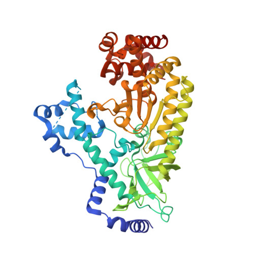
Ligand-induced expansion of the S1' site in the anthrax toxin lethal factor.

Maize, K.M.Kurbanov, E.K.Johnson, R.L.Amin, E.A.Finzel, B.C.

(2015) FEBS Lett 589: 3836-3841

  • DOI: https://doi.org/10.1016/j.febslet.2015.11.005
  • Primary Citation of Related Structures:  
    4XM6, 4XM7, 4XM8

  • PubMed Abstract: 

    The Bacillus anthracis lethal factor (LF) is one component of a tripartite exotoxin partly responsible for persistent anthrax cytotoxicity after initial bacterial infection. Inhibitors of the zinc metalloproteinase have been investigated as potential therapeutic agents, but LF is a challenging target because inhibitors lack sufficient selectivity or possess poor pharmaceutical properties. These structural studies reveal an alternate conformation of the enzyme, induced upon binding of specific inhibitors, that opens a previously unobserved deep pocket termed S1'(∗) which might afford new opportunities to design selective inhibitors that target this subsite.


  • Organizational Affiliation

    Department of Medicinal Chemistry, University of Minnesota, 308 Harvard St SE, 8-101 Weaver-Densford Hall, Minneapolis, MN 55455, United States.


Macromolecules
Find similar proteins by:  (by identity cutoff)  |  3D Structure
Entity ID: 1
MoleculeChains Sequence LengthOrganismDetailsImage
Lethal factor519Bacillus anthracisMutation(s): 1 
Gene Names: lefpXO1-107BXA0172GBAA_pXO1_0172
EC: 3.4.24.83
UniProt
Find proteins for P15917 (Bacillus anthracis)
Explore P15917 
Go to UniProtKB:  P15917
Entity Groups  
Sequence Clusters30% Identity50% Identity70% Identity90% Identity95% Identity100% Identity
UniProt GroupP15917
Sequence Annotations
Expand
  • Reference Sequence
Experimental Data & Validation

Experimental Data

  • Method: X-RAY DIFFRACTION
  • Resolution: 2.35 Å
  • R-Value Free:  0.246 (Depositor), 0.250 (DCC) 
  • R-Value Work:  0.205 (Depositor), 0.210 (DCC) 
  • R-Value Observed: 0.207 (Depositor) 
Space Group: P 21 21 21
Unit Cell:
Length ( Å )Angle ( ˚ )
a = 57.936α = 90
b = 76.922β = 90
c = 138.576γ = 90
Software Package:
Software NamePurpose
HKL-2000data scaling
HKL-2000data reduction
PHASERphasing
PHENIXrefinement
PDB_EXTRACTdata extraction

Structure Validation

View Full Validation Report


Ligand Structure Quality Assessment 

Created with Raphaël 2.3.0Worse 01 BetterLigand structure goodness of fit to experimental dataBest fitted 41RClick on this verticalbar to view details

Entry History & Funding Information

Deposition Data


Funding OrganizationLocationGrant Number
National Institutes of Health/National Institute Of Allergy and Infectious Diseases (NIH/NIAID)United StatesAI091790-01
Minnesota Dept. of Employment and Economic DevelopmentUnited StatesSPAP-06-0014-P-FY07

Revision History  (Full details and data files)

  • Version 1.0: 2015-11-11
    Type: Initial release
  • Version 1.1: 2015-12-02
    Changes: Database references
  • Version 1.2: 2015-12-30
    Changes: Database references
  • Version 1.3: 2017-09-27
    Changes: Author supporting evidence, Database references, Derived calculations
  • Version 1.4: 2019-12-11
    Changes: Author supporting evidence
  • Version 1.5: 2023-09-27
    Changes: Data collection, Database references, Refinement description