4XKC | pdb_00004xkc

Crystal Structure of GENOMES UNCOUPLED 4 (GUN4) in Complex with Magnesium Deuteroporphyrin IX


Experimental Data Snapshot

  • Method: X-RAY DIFFRACTION
  • Resolution: 2.00 Å
  • R-Value Free: 
    0.229 (Depositor), 0.228 (DCC) 
  • R-Value Work: 
    0.182 (Depositor) 
  • R-Value Observed: 
    0.184 (Depositor) 

wwPDB Validation 3D Report Full Report

Validation slider image for 4XKC

Ligand Structure Quality Assessment 


This is version 1.3 of the entry. See complete history

Literature

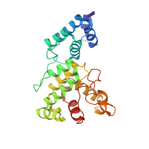
Crystal Structures of GUN4 in Complex with Porphyrins.

Chen, X.Pu, H.Wang, X.Long, W.Lin, R.Liu, L.

(2015) Mol Plant 8: 1125-1127

  • DOI: https://doi.org/10.1016/j.molp.2015.04.013
  • Primary Citation Related Structures: 
    4XKB, 4XKC

  • Organizational Affiliation
    • Photosynthesis Research Center, Key Laboratory of Photobiology, Institute of Botany, Chinese Academy of Sciences, 20 Nanxincun, Haidian District, Beijing 100093, China; University of Chinese Academy of Sciences, 19A Yuquan Road, Haidian District, Beijing 100049, China.

Macromolecule Content 

  • Total Structure Weight: 27.02 kDa 
  • Atom Count: 2,121 
  • Modeled Residue Count: 225 
  • Deposited Residue Count: 233 
  • Unique protein chains: 1

Macromolecules

Find similar proteins by:|  3D Structure
Entity ID: 1
MoleculeChains  Sequence LengthOrganismDetailsImage
Ycf53-like protein233Synechocystis sp. PCC 6803 substr. KazusaMutation(s): 0 
Gene Names: sll0558
UniProt
Find proteins for P72583 (Synechocystis sp. (strain ATCC 27184 / PCC 6803 / Kazusa))
Explore P72583 
Go to UniProtKB:  P72583
Entity Groups
Sequence Clusters30% Identity50% Identity70% Identity90% Identity95% Identity100% Identity
UniProt GroupP72583
Sequence Annotations
Expand
Reference Sequence

Small Molecules

Ligands 1 Unique
IDChains Name / Formula / InChI Key2D Diagram3D Interactions
MD9

Query on MD9


Download:Ideal Coordinates CCD File
B [auth A]Magnesium deuteroporphyrin IX
C30 H28 Mg N4 O4
WOJNEAWSXKLRSI-RWRCOHKGSA-L

Experimental Data & Validation

Experimental Data

  • Method: X-RAY DIFFRACTION
  • Resolution: 2.00 Å
  • R-Value Free:  0.229 (Depositor), 0.228 (DCC) 
  • R-Value Work:  0.182 (Depositor) 
  • R-Value Observed: 0.184 (Depositor) 
Space Group: P 21 21 21
Unit Cell:
Length ( Å )Angle ( ˚ )
a = 32.849α = 90
b = 64.026β = 90
c = 106.144γ = 90
Software Package:
Software NamePurpose
HKL-2000refinement
HKL-2000data reduction
HKL-2000data scaling
PHASERphasing
PHENIXrefinement

Structure Validation

View Full Validation Report


Ligand Structure Quality Assessment 


Entry History 

Deposition Data

Revision History  (Full details and data files)

  • Version 1.0: 2015-05-20
    Type: Initial release
  • Version 1.1: 2015-07-15
    Changes: Database references
  • Version 1.2: 2015-07-22
    Changes: Database references
  • Version 1.3: 2024-03-20
    Changes: Data collection, Database references, Derived calculations, Source and taxonomy, Structure summary