4XJR | pdb_00004xjr

The catalytic mechanism of human parainfluenza virus type 3 haemagglutinin-neuraminidase revealed


Experimental Data Snapshot

  • Method: X-RAY DIFFRACTION
  • Resolution: 3.00 Å
  • R-Value Free: 
    0.298 (Depositor) 
  • R-Value Work: 
    0.190 (Depositor) 
  • R-Value Observed: 
    0.195 (Depositor) 

wwPDB Validation 3D Report Full Report

Validation slider image for 4XJR

Ligand Structure Quality Assessment 


This is version 3.1 of the entry. See complete history

Literature

The catalytic mechanism of human parainfluenza virus type 3 haemagglutinin-neuraminidase revealed.

Dirr, L.El-Deeb, I.M.Guillon, P.Carroux, C.J.Chavas, L.M.von Itzstein, M.

(2015) Angew Chem Int Ed Engl 54: 2936-2940

  • DOI: https://doi.org/10.1002/anie.201412243
  • Primary Citation Related Structures: 
    4XJQ, 4XJR

  • PubMed Abstract: 

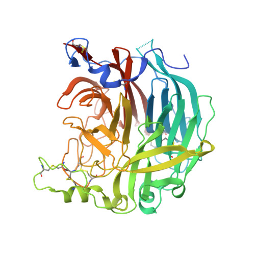
    Human parainfluenza virus type 3 (hPIV-3) is one of the leading causes for lower respiratory tract disease in children, with neither an approved antiviral drug nor vaccine available to date. Understanding the catalytic mechanism of human parainfluenza virus haemagglutinin-neuraminidase (HN) protein is key to the design of specific inhibitors against this virus. Herein, we used (1) H NMR spectroscopy, X-ray crystallography, and virological assays to study the catalytic mechanism of the HN enzyme activity and have identified the conserved Tyr530 as a key amino acid involved in catalysis. A novel 2,3-difluorosialic acid derivative showed prolonged enzyme inhibition and was found to react and form a covalent bond with Tyr530. Furthermore, the novel derivative exhibited enhanced potency in virus blockade assays relative to its Neu2en analogue. These outcomes open the door for a new generation of potent inhibitors against hPIV-3 HN.


  • Organizational Affiliation
    • Institute for Glycomics, Gold Coast Campus, Griffith University, Queensland, 4222 (Australia).

Macromolecule Content 

  • Total Structure Weight: 104.23 kDa 
  • Atom Count: 6,869 
  • Modeled Residue Count: 855 
  • Deposited Residue Count: 908 
  • Unique protein chains: 1

Macromolecules

Find similar proteins by:|  3D Structure
Entity ID: 1
MoleculeChains  Sequence LengthOrganismDetailsImage
Hemagglutinin-neuraminidase
A, B
454Human respirovirus 3Mutation(s): 0 
EC: 3.2.1.18
UniProt
Find proteins for P08492 (Human parainfluenza 3 virus (strain Wash/47885/57))
Explore P08492 
Go to UniProtKB:  P08492
Entity Groups
Sequence Clusters30% Identity50% Identity70% Identity90% Identity95% Identity100% Identity
UniProt GroupP08492
Glycosylation
Glycosylation Sites: 2
Sequence Annotations
Expand
Reference Sequence

Oligosaccharides

Help  
Entity ID: 2
MoleculeChains Length2D Diagram GlycosylationD Interactions
2-acetamido-2-deoxy-beta-D-glucopyranose-(1-4)-2-acetamido-2-deoxy-beta-D-glucopyranose
C
2N-Glycosylation
Glycosylation Resources
GlyTouCan: G42666HT
GlyCosmos: G42666HT
GlyGen: G42666HT
Entity ID: 3
MoleculeChains Length2D Diagram GlycosylationD Interactions
beta-D-mannopyranose-(1-3)-2-acetamido-2-deoxy-beta-D-glucopyranose-(1-4)-2-acetamido-2-deoxy-beta-D-glucopyranose
D
3N-Glycosylation
Glycosylation Resources
GlyTouCan: G44266EX
GlyCosmos: G44266EX
GlyGen: G44266EX

Small Molecules

Ligands 5 Unique
IDChains Name / Formula / InChI Key2D Diagram3D Interactions
OEL

Query on OEL


Download:Ideal Coordinates CCD File
H [auth A],
K [auth B]
(6R)-2,6-anhydro-3,4,5-trideoxy-6-[(2S)-2,3-dihydroxypropanoyl]-3-fluoro-5-[(2-methylpropanoyl)amino]-4-triaza-1,2-dien -2-ium-1-yl-L-gulonic acid
C13 H20 F N4 O7
UFSBEYSJWUBWLQ-JAYUSQFOSA-O
NAG

Query on NAG


Download:Ideal Coordinates CCD File
G [auth A],
J [auth B]
2-acetamido-2-deoxy-beta-D-glucopyranose
C8 H15 N O6
OVRNDRQMDRJTHS-FMDGEEDCSA-N
SO4

Query on SO4


Download:Ideal Coordinates CCD File
F [auth A]SULFATE ION
O4 S
QAOWNCQODCNURD-UHFFFAOYSA-L
EDO

Query on EDO


Download:Ideal Coordinates CCD File
L [auth B]1,2-ETHANEDIOL
C2 H6 O2
LYCAIKOWRPUZTN-UHFFFAOYSA-N
CA

Query on CA


Download:Ideal Coordinates CCD File
E [auth A],
I [auth B]
CALCIUM ION
Ca
BHPQYMZQTOCNFJ-UHFFFAOYSA-N

Experimental Data & Validation

Experimental Data

  • Method: X-RAY DIFFRACTION
  • Resolution: 3.00 Å
  • R-Value Free:  0.298 (Depositor) 
  • R-Value Work:  0.190 (Depositor) 
  • R-Value Observed: 0.195 (Depositor) 
Space Group: P 21 21 21
Unit Cell:
Length ( Å )Angle ( ˚ )
a = 81.85α = 90
b = 98.303β = 90
c = 103.261γ = 90
Software Package:
Software NamePurpose
REFMACrefinement
HKL-2000data collection
XDSdata reduction
Aimlessdata scaling
PHASERphasing
PDB_EXTRACTdata extraction

Structure Validation

View Full Validation Report


Ligand Structure Quality Assessment 


Entry History 

& Funding Information

Deposition Data


Funding OrganizationLocationGrant Number
Australian Research Council (ARC)AustraliaDP1094549
National Health and Medical Research Council (NHMRC, Australia)AustraliaID1047824

Revision History  (Full details and data files)

  • Version 1.0: 2015-02-11
    Type: Initial release
  • Version 1.1: 2015-03-04
    Changes: Database references
  • Version 2.0: 2017-09-06
    Changes: Atomic model, Author supporting evidence, Data collection, Database references, Derived calculations, Source and taxonomy, Structure summary
  • Version 2.1: 2020-01-01
    Changes: Author supporting evidence
  • Version 3.0: 2020-07-29
    Type: Remediation
    Reason: Carbohydrate remediation
    Changes: Advisory, Atomic model, Data collection, Derived calculations, Structure summary
  • Version 3.1: 2024-11-06
    Changes: Data collection, Database references, Derived calculations, Structure summary