4XHZ | pdb_00004xhz

Crystal Structure of Human Protocadherin-15 EC8-10


Experimental Data Snapshot

  • Method: X-RAY DIFFRACTION
  • Resolution: 2.80 Å
  • R-Value Free: 
    0.200 (Depositor), 0.215 (DCC) 
  • R-Value Work: 
    0.170 (Depositor) 
  • R-Value Observed: 
    0.171 (Depositor) 

Starting Models: experimental
View more details

wwPDB Validation 3D Report Full Report

Validation slider image for 4XHZ

This is version 1.5 of the entry. See complete history

Literature

An elastic element in the protocadherin-15 tip link of the inner ear.

Araya-Secchi, R.Neel, B.L.Sotomayor, M.

(2016) Nat Commun 7: 13458-13458

  • DOI: https://doi.org/10.1038/ncomms13458
  • Primary Citation Related Structures: 
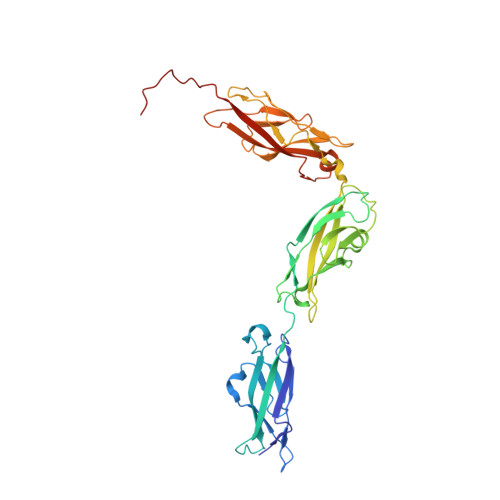
    4XHZ, 5KJ4

  • PubMed Abstract: 

    Tip link filaments convey force and gate inner-ear hair-cell transduction channels to mediate perception of sound and head movements. Cadherin-23 and protocadherin-15 form tip links through a calcium-dependent interaction of their extracellular domains made of multiple extracellular cadherin (EC) repeats. These repeats are structurally similar, but not identical in sequence, often featuring linkers with conserved calcium-binding sites that confer mechanical strength to them. Here we present the X-ray crystal structures of human protocadherin-15 EC8-EC10 and mouse EC9-EC10, which show an EC8-9 canonical-like calcium-binding linker, and an EC9-10 calcium-free linker that alters the linear arrangement of EC repeats. Molecular dynamics simulations and small-angle X-ray scattering experiments support this non-linear conformation. Simulations also suggest that unbending of EC9-10 confers some elasticity to otherwise rigid tip links. The new structure provides a first view of protocadherin-15's non-canonical EC linkers and suggests how they may function in inner-ear mechanotransduction, with implications for other cadherins.


  • Organizational Affiliation
    • Department of Chemistry and Biochemistry, The Ohio State University, 484 W 12th Avenue, Columbus, Ohio 43210, USA.

Macromolecule Content 

  • Total Structure Weight: 37.59 kDa 
  • Atom Count: 2,664 
  • Modeled Residue Count: 333 
  • Deposited Residue Count: 338 
  • Unique protein chains: 1

Macromolecules

Find similar proteins by:|  3D Structure
Entity ID: 1
MoleculeChains  Sequence LengthOrganismDetailsImage
Protocadherin-15338Homo sapiensMutation(s): 0 
Gene Names: PCDH15USH1F
UniProt & NIH Common Fund Data Resources
Find proteins for Q96QU1 (Homo sapiens)
Explore Q96QU1 
Go to UniProtKB:  Q96QU1
PHAROS:  Q96QU1
GTEx:  ENSG00000150275 
Entity Groups
Sequence Clusters30% Identity50% Identity70% Identity90% Identity95% Identity100% Identity
UniProt GroupQ96QU1
Sequence Annotations
Expand
Reference Sequence

Experimental Data & Validation

Experimental Data

  • Method: X-RAY DIFFRACTION
  • Resolution: 2.80 Å
  • R-Value Free:  0.200 (Depositor), 0.215 (DCC) 
  • R-Value Work:  0.170 (Depositor) 
  • R-Value Observed: 0.171 (Depositor) 
Space Group: P 63 2 2
Unit Cell:
Length ( Å )Angle ( ˚ )
a = 157.929α = 90
b = 157.929β = 90
c = 142.898γ = 120
Software Package:
Software NamePurpose
REFMACrefinement
HKL-2000data reduction
HKL-2000data scaling
PHASERphasing

Structure Validation

View Full Validation Report


Entry History 

& Funding Information

Deposition Data


Funding OrganizationLocationGrant Number
National Institutes of Health/National Institute on Deafness and Other Communication Disorders (NIH/NIDCD)United StatesK99/R00 DC012534

Revision History  (Full details and data files)

  • Version 1.0: 2016-09-21
    Type: Initial release
  • Version 1.1: 2016-11-23
    Changes: Database references
  • Version 1.2: 2016-12-21
    Changes: Database references
  • Version 1.3: 2017-09-06
    Changes: Author supporting evidence
  • Version 1.4: 2019-12-18
    Changes: Author supporting evidence
  • Version 1.5: 2023-09-27
    Changes: Data collection, Database references, Derived calculations, Refinement description