4XDA | pdb_00004xda

Vibrio cholerae O395 Ribokinase complexed with Ribose, ADP and Sodium ion.


Experimental Data Snapshot

  • Method: X-RAY DIFFRACTION
  • Resolution: 1.75 Å
  • R-Value Free: 
    0.225 (Depositor), 0.225 (DCC) 
  • R-Value Work: 
    0.186 (Depositor), 0.190 (DCC) 
  • R-Value Observed: 
    0.189 (Depositor) 

wwPDB Validation 3D Report Full Report

Validation slider image for 4XDA

Ligand Structure Quality Assessment 


This is version 2.1 of the entry. See complete history

Literature

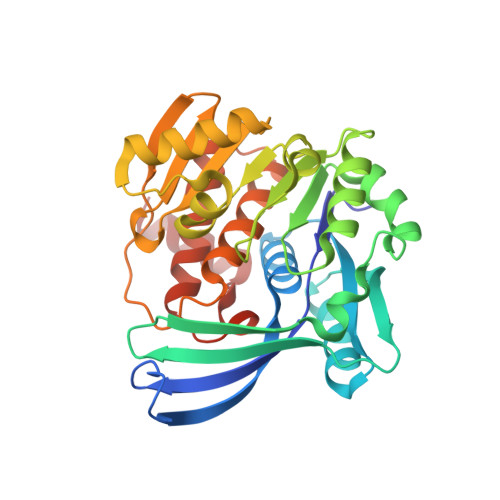
Crystal Structure of Apo and Ligand Bound Vibrio cholerae Ribokinase (Vc-RK): Role of Monovalent Cation Induced Activation and Structural Flexibility in Sugar Phosphorylation

Paul, R.Patra, M.D.Sen, U.

(2015) Adv Exp Med Biol 842: 293-307

Macromolecule Content 

  • Total Structure Weight: 132.25 kDa 
  • Atom Count: 10,337 
  • Modeled Residue Count: 1,224 
  • Deposited Residue Count: 1,236 
  • Unique protein chains: 1

Macromolecules

Find similar proteins by:|  3D Structure
Entity ID: 1
MoleculeChains  Sequence LengthOrganismDetailsImage
Ribokinase
A, B, C, D
309Vibrio cholerae O395Mutation(s): 0 
Gene Names: rbsKVC0395_0007VC395_A0124
EC: 2.7.1.15
UniProt
Find proteins for A0A0H2UL04 (Vibrio cholerae serotype O1 (strain ATCC 39541 / Classical Ogawa 395 / O395))
Explore A0A0H2UL04 
Go to UniProtKB:  A0A0H2UL04
Entity Groups
Sequence Clusters30% Identity50% Identity70% Identity90% Identity95% Identity100% Identity
UniProt GroupA0A0H2UL04
Sequence Annotations
Expand
Reference Sequence

Small Molecules

Ligands 3 Unique
IDChains Name / Formula / InChI Key2D Diagram3D Interactions
ADP

Query on ADP


Download:Ideal Coordinates CCD File
F [auth A],
H [auth B],
I [auth B],
L [auth C],
O [auth D]
ADENOSINE-5'-DIPHOSPHATE
C10 H15 N5 O10 P2
XTWYTFMLZFPYCI-KQYNXXCUSA-N
RIB

Query on RIB


Download:Ideal Coordinates CCD File
E [auth A],
K [auth C],
N [auth D]
alpha-D-ribofuranose
C5 H10 O5
HMFHBZSHGGEWLO-AIHAYLRMSA-N
NA

Query on NA


Download:Ideal Coordinates CCD File
G [auth A],
J [auth B],
M [auth C],
P [auth D]
SODIUM ION
Na
FKNQFGJONOIPTF-UHFFFAOYSA-N

Experimental Data & Validation

Experimental Data

  • Method: X-RAY DIFFRACTION
  • Resolution: 1.75 Å
  • R-Value Free:  0.225 (Depositor), 0.225 (DCC) 
  • R-Value Work:  0.186 (Depositor), 0.190 (DCC) 
  • R-Value Observed: 0.189 (Depositor) 
Space Group: P 1
Unit Cell:
Length ( Å )Angle ( ˚ )
a = 59.42α = 106.29
b = 70.7β = 97.65
c = 79.88γ = 98.68
Software Package:
Software NamePurpose
PHENIXrefinement
MOSFLMdata reduction
SCALAdata scaling
PHASERphasing

Structure Validation

View Full Validation Report


Ligand Structure Quality Assessment 


Entry History 

& Funding Information

Deposition Data


Funding OrganizationLocationGrant Number
Department of Biotechnology, Department of Atomic EnergyIndiaMSACR project

Revision History  (Full details and data files)

  • Version 1.0: 2015-03-04
    Type: Initial release
  • Version 2.0: 2020-07-29
    Type: Remediation
    Reason: Carbohydrate remediation
    Changes: Atomic model, Data collection, Database references, Derived calculations, Source and taxonomy, Structure summary
  • Version 2.1: 2024-03-20
    Changes: Data collection, Database references, Structure summary