4XBM | pdb_00004xbm

X-ray crystal structure of Notch ligand Delta-like 1


Experimental Data Snapshot

  • Method: X-RAY DIFFRACTION
  • Resolution: 3.20 Å
  • R-Value Free: 
    0.283 (Depositor), 0.289 (DCC) 
  • R-Value Work: 
    0.258 (Depositor), 0.263 (DCC) 
  • R-Value Observed: 
    0.260 (Depositor) 

Starting Model: experimental
View more details

wwPDB Validation 3D Report Full Report

Validation slider image for 4XBM

Ligand Structure Quality Assessment 


This is version 1.7 of the entry. See complete history

Literature

Notch ligand delta-like1: X-ray crystal structure and binding affinity.

Kershaw, N.J.Church, N.L.Griffin, M.D.Luo, C.S.Adams, T.E.Burgess, A.W.

(2015) Biochem J 468: 159-166

  • DOI: https://doi.org/10.1042/BJ20150010
  • Primary Citation Related Structures: 
    4XBM

  • PubMed Abstract: 

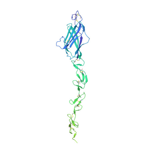
    The Notch pathway is a fundamental signalling system in most multicellular animals. We have determined the X-ray crystal structure of the extracellular domain of the Notch ligand delta-like ligand-1 (Dll-1). The structure incorporates the N-terminal C2 domain, receptor-binding DSL domain and the first six (of eight) EGF (epidermal growth factor)-like repeats, which form a highly extended conformation, confirmed by analytical ultracentrifugation. Comparison of our structure with a fragment of Jagged1 ligand allows us to dissect the similarities and differences between the ligand families. Differences in the C2 domains of Dll-1 and Jagged1 suggest their lipid-binding properties are likely to differ. A conserved hydrophobic patch on the surface of both Dll-1 and Jagged1 provides a likely receptor-interaction site that is common to both ligands. We also explore the binding affinity of Dll-1 for a fragment of Notch1 using different techniques. Apparent binding affinities vary when different techniques are used, explaining discrepancies in the literature. Using analytical ultracentrifugation, we perform for the first time binding analyses where both receptor and ligand are in solution, which confirms a Kd of 10 μM for this interaction.


  • Organizational Affiliation
    • *Department of Structural Biology, Walter and Eliza Hall Institute of Medical Research and Department of Medical Biology, University of Melbourne, Parkville, 3052 Australia.

Macromolecule Content 

  • Total Structure Weight: 115.03 kDa 
  • Atom Count: 4,996 
  • Modeled Residue Count: 654 
  • Deposited Residue Count: 1,062 
  • Unique protein chains: 1

Macromolecules

Find similar proteins by:|  3D Structure
Entity ID: 1
MoleculeChains  Sequence LengthOrganismDetailsImage
Delta-like protein 1
A, B
531Homo sapiensMutation(s): 0 
Gene Names: DLL1UNQ146/PRO172
UniProt & NIH Common Fund Data Resources
Find proteins for O00548 (Homo sapiens)
Explore O00548 
Go to UniProtKB:  O00548
PHAROS:  O00548
GTEx:  ENSG00000198719 
Entity Groups
Sequence Clusters30% Identity50% Identity70% Identity90% Identity95% Identity100% Identity
UniProt GroupO00548
Glycosylation
Glycosylation Sites: 1Go to GlyGen: O00548-1
Sequence Annotations
Expand
Reference Sequence

Small Molecules

Ligands 1 Unique
IDChains Name / Formula / InChI Key2D Diagram3D Interactions
FUC

Query on FUC


Download:Ideal Coordinates CCD File
C [auth B]alpha-L-fucopyranose
C6 H12 O5
SHZGCJCMOBCMKK-SXUWKVJYSA-N

Experimental Data & Validation

Experimental Data

  • Method: X-RAY DIFFRACTION
  • Resolution: 3.20 Å
  • R-Value Free:  0.283 (Depositor), 0.289 (DCC) 
  • R-Value Work:  0.258 (Depositor), 0.263 (DCC) 
  • R-Value Observed: 0.260 (Depositor) 
Space Group: P 21 21 21
Unit Cell:
Length ( Å )Angle ( ˚ )
a = 108.15α = 90
b = 118.87β = 90
c = 134.81γ = 90
Software Package:
Software NamePurpose
PHENIXrefinement
XDSdata reduction
PHASERphasing
PDB_EXTRACTdata extraction
XSCALEdata scaling

Structure Validation

View Full Validation Report


Ligand Structure Quality Assessment 


Entry History 

& Funding Information

Deposition Data


Funding OrganizationLocationGrant Number
National Health and Medical Research Council (NHMRC, Australia)Australia487922

Revision History  (Full details and data files)

  • Version 1.0: 2015-03-11
    Type: Initial release
  • Version 1.1: 2015-05-20
    Changes: Database references
  • Version 1.2: 2015-05-27
    Changes: Data collection, Structure summary
  • Version 1.3: 2017-09-06
    Changes: Author supporting evidence, Data collection, Database references, Derived calculations, Source and taxonomy, Structure summary
  • Version 1.4: 2020-01-08
    Changes: Author supporting evidence, Data collection
  • Version 1.5: 2020-07-29
    Type: Remediation
    Reason: Carbohydrate remediation
    Changes: Data collection, Derived calculations, Structure summary
  • Version 1.6: 2023-09-27
    Changes: Data collection, Database references, Refinement description, Structure summary
  • Version 1.7: 2024-10-23
    Changes: Structure summary