4X8T | pdb_00004x8t

FACTOR VIIA IN COMPLEX WITH THE INHIBITOR 7-chloro-3,4-dihydroisoquinolin-1(2H)-one


Experimental Data Snapshot

  • Method: X-RAY DIFFRACTION
  • Resolution: 2.20 Å
  • R-Value Free: 
    0.221 (Depositor), 0.222 (DCC) 
  • R-Value Work: 
    0.198 (Depositor), 0.195 (DCC) 
  • R-Value Observed: 
    0.200 (Depositor) 

Starting Model: experimental
View more details

wwPDB Validation 3D Report Full Report

Validation slider image for 4X8T

Ligand Structure Quality Assessment 


This is version 1.2 of the entry. See complete history

Literature

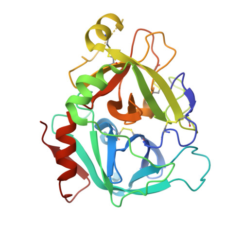
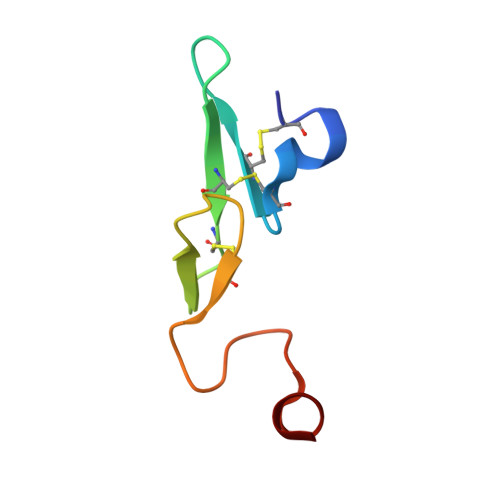
Discovery of Novel P1 Groups for Coagulation Factor VIIa Inhibition Using Fragment-Based Screening.

Cheney, D.L.Bozarth, J.M.Metzler, W.J.Morin, P.E.Mueller, L.Newitt, J.A.Nirschl, A.H.Rendina, A.R.Tamura, J.K.Wei, A.Wen, X.Wurtz, N.R.Seiffert, D.A.Wexler, R.R.Priestley, E.S.

(2015) J Med Chem 58: 2799-2808

  • DOI: https://doi.org/10.1021/jm501982k
  • Primary Citation Related Structures: 
    4X8S, 4X8T, 4X8U, 4X8V

  • PubMed Abstract: 

    A multidisciplinary, fragment-based screening approach involving protein ensemble docking and biochemical and NMR assays is described. This approach led to the discovery of several structurally diverse, neutral surrogates for cationic factor VIIa P1 groups, which are generally associated with poor pharmacokinetic (PK) properties. Among the novel factor VIIa inhibitory fragments identified were aryl halides, lactams, and heterocycles. Crystallographic structures for several bound fragments were obtained, leading to the successful design of a potent factor VIIa inhibitor with a neutral lactam P1 and improved permeability.


  • Organizational Affiliation
    • †Bristol-Myers Squibb Co., Research and Development, 311 Pennington-Rocky Hill Road, Pennington, New Jersey 08543, United States.

Macromolecule Content 

  • Total Structure Weight: 34.74 kDa 
  • Atom Count: 2,502 
  • Modeled Residue Count: 305 
  • Deposited Residue Count: 309 
  • Unique protein chains: 2

Macromolecules

Find similar proteins by:|  3D Structure
Entity ID: 1
MoleculeChains  Sequence LengthOrganismDetailsImage
Factor VIIa (Heavy Chain)A [auth H]254Homo sapiensMutation(s): 0 
Gene Names: F7
EC: 3.4.21.21
UniProt & NIH Common Fund Data Resources
Find proteins for P08709 (Homo sapiens)
Explore P08709 
Go to UniProtKB:  P08709
PHAROS:  P08709
GTEx:  ENSG00000057593 
Entity Groups
Sequence Clusters30% Identity50% Identity70% Identity90% Identity95% Identity100% Identity
UniProt GroupP08709
Sequence Annotations
Expand
Reference Sequence
Find similar proteins by:|  3D Structure
Entity ID: 2
MoleculeChains  Sequence LengthOrganismDetailsImage
Factor VIIa (Light Chain)B [auth L]55Homo sapiensMutation(s): 0 
Gene Names: F7
EC: 3.4.21.21
UniProt & NIH Common Fund Data Resources
Find proteins for P08709 (Homo sapiens)
Explore P08709 
Go to UniProtKB:  P08709
PHAROS:  P08709
GTEx:  ENSG00000057593 
Entity Groups
Sequence Clusters30% Identity50% Identity70% Identity90% Identity95% Identity100% Identity
UniProt GroupP08709
Sequence Annotations
Expand
Reference Sequence

Small Molecules

Ligands 4 Unique
IDChains Name / Formula / InChI Key2D Diagram3D Interactions
3Z8

Query on 3Z8


Download:Ideal Coordinates CCD File
C [auth H]7-chloro-3,4-dihydroisoquinolin-1(2H)-one
C9 H8 Cl N O
NMZRTRAYSHQMPR-UHFFFAOYSA-N
SO4

Query on SO4


Download:Ideal Coordinates CCD File
E [auth H],
F [auth H],
G [auth H]
SULFATE ION
O4 S
QAOWNCQODCNURD-UHFFFAOYSA-L
GOL

Query on GOL


Download:Ideal Coordinates CCD File
H
GLYCEROL
C3 H8 O3
PEDCQBHIVMGVHV-UHFFFAOYSA-N
CA

Query on CA


Download:Ideal Coordinates CCD File
D [auth H]CALCIUM ION
Ca
BHPQYMZQTOCNFJ-UHFFFAOYSA-N
Binding Affinity Annotations 
IDSourceBinding Affinity
3Z8 BindingDB:  4X8T Ki: 8.90e+6 (nM) from 1 assay(s)

Experimental Data & Validation

Experimental Data

  • Method: X-RAY DIFFRACTION
  • Resolution: 2.20 Å
  • R-Value Free:  0.221 (Depositor), 0.222 (DCC) 
  • R-Value Work:  0.198 (Depositor), 0.195 (DCC) 
  • R-Value Observed: 0.200 (Depositor) 
Space Group: P 41 21 2
Unit Cell:
Length ( Å )Angle ( ˚ )
a = 94.48α = 90
b = 94.48β = 90
c = 108.09γ = 90
Software Package:
Software NamePurpose
BUSTER-TNTrefinement
PDB_EXTRACTdata extraction
HKL-2000data reduction
HKL-2000data scaling
AMoREphasing

Structure Validation

View Full Validation Report


Ligand Structure Quality Assessment 


Entry History 

Deposition Data

  • Released Date: 2015-03-25 
  • Deposition Author(s): Wei, A.

Revision History  (Full details and data files)

  • Version 1.0: 2015-03-25
    Type: Initial release
  • Version 1.1: 2015-04-08
    Changes: Database references
  • Version 1.2: 2024-11-06
    Changes: Data collection, Database references, Derived calculations, Refinement description, Source and taxonomy, Structure summary