4X7Z

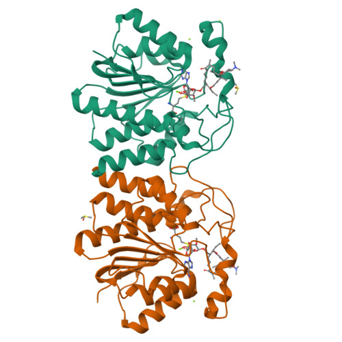
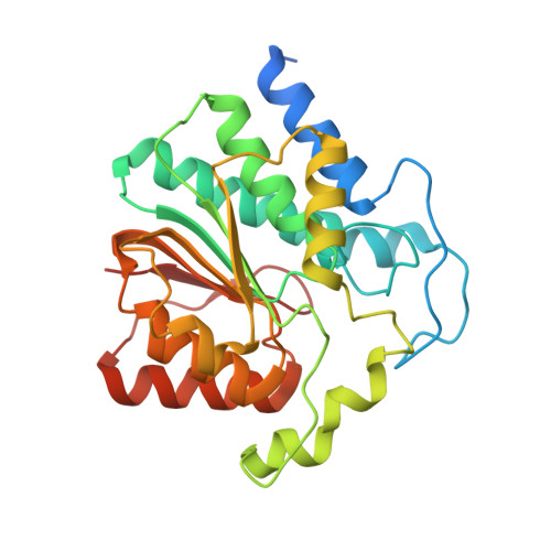
MycF mycinamicin III 3'-O-methyltransferase (E35Q, M56A, E139A variant) in complex with Mg, SAH and mycinamicin III (substrate)


Experimental Data Snapshot

  • Method: X-RAY DIFFRACTION
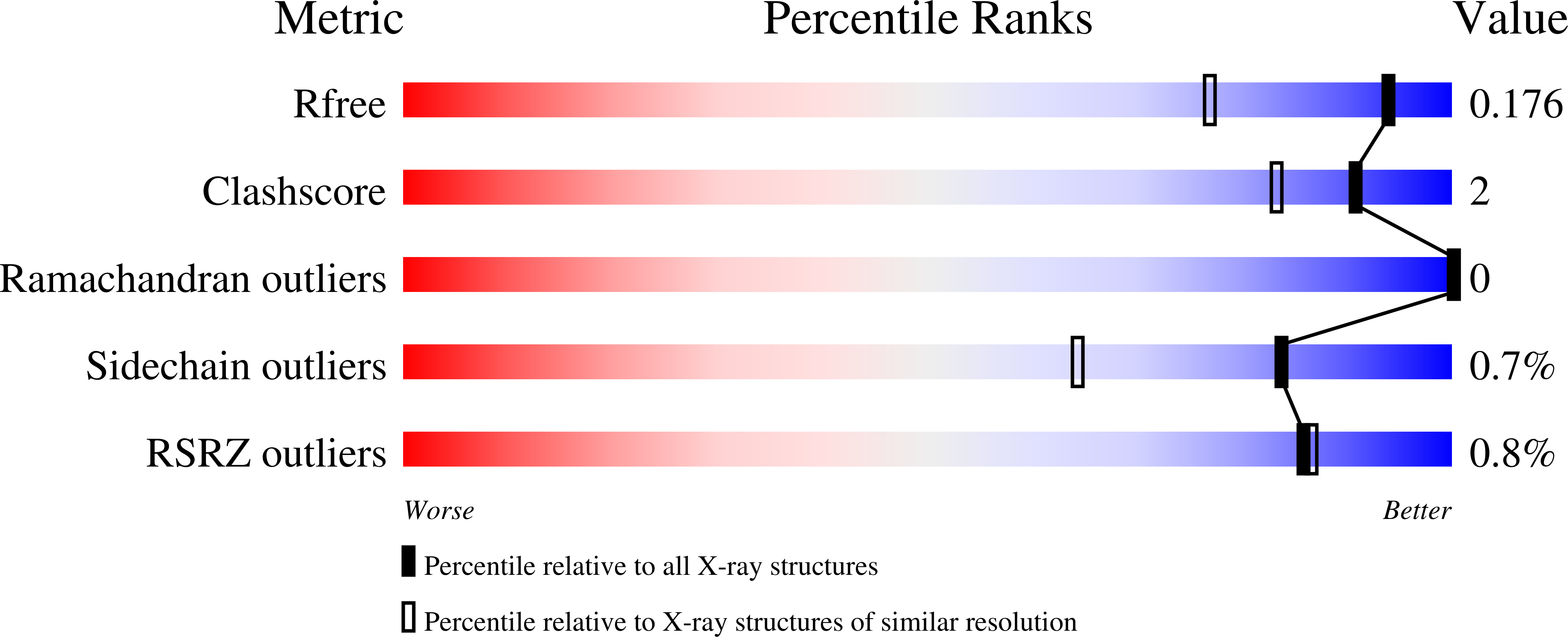
  • Resolution: 1.44 Å
  • R-Value Free: 0.168 
  • R-Value Work: 0.153 
  • R-Value Observed: 0.154 

wwPDB Validation   3D Report Full Report


Ligand Structure Quality Assessment 

Created with Raphaël 2.3.0Worse 01 BetterLigand structure goodness of fit to experimental dataBest fitted ZM3Click on this verticalbar to view detailsBest fitted SAHClick on this verticalbar to view details

This is version 1.3 of the entry. See complete history


Literature

Structural Basis of Substrate Specificity and Regiochemistry in the MycF/TylF Family of Sugar O-Methyltransferases.

Bernard, S.M.Akey, D.L.Tripathi, A.Park, S.R.Konwerski, J.R.Anzai, Y.Li, S.Kato, F.Sherman, D.H.Smith, J.L.

(2015) ACS Chem Biol 10: 1340-1351

  • DOI: https://doi.org/10.1021/cb5009348
  • Primary Citation of Related Structures:  
    4X7U, 4X7V, 4X7W, 4X7X, 4X7Y, 4X7Z, 4X81, 4XVY, 4XVZ

  • PubMed Abstract: 

    Sugar moieties in natural products are frequently modified by O-methylation. In the biosynthesis of the macrolide antibiotic mycinamicin, methylation of a 6'-deoxyallose substituent occurs in a stepwise manner first at the 2'- and then the 3'-hydroxyl groups to produce the mycinose moiety in the final product. The timing and placement of the O-methylations impact final stage C-H functionalization reactions mediated by the P450 monooxygenase MycG. The structural basis of pathway ordering and substrate specificity is unknown. A series of crystal structures of MycF, the 3'-O-methyltransferase, including the free enzyme and complexes with S-adenosyl homocysteine (SAH), substrate, product, and unnatural substrates, show that SAM binding induces substantial ordering that creates the binding site for the natural substrate, and a bound metal ion positions the substrate for catalysis. A single amino acid substitution relaxed the 2'-methoxy specificity but retained regiospecificity. The engineered variant produced a new mycinamicin analog, demonstrating the utility of structural information to facilitate bioengineering approaches for the chemoenzymatic synthesis of complex small molecules containing modified sugars. Using the MycF substrate complex and the modeled substrate complex of a 4'-specific homologue, active site residues were identified that correlate with the 3' or 4' specificity of MycF family members and define the protein and substrate features that direct the regiochemistry of methyltransfer. This classification scheme will be useful in the annotation of new secondary metabolite pathways that utilize this family of enzymes.


  • Organizational Affiliation

    †Chemical Biology Doctoral Program, University of Michigan, Ann Arbor, Michigan 48109, United States.


Macromolecules
Find similar proteins by:  (by identity cutoff)  |  3D Structure
Entity ID: 1
MoleculeChains Sequence LengthOrganismDetailsImage
Mycinamicin III 3''-O-methyltransferase
A, B
274Micromonospora griseorubidaMutation(s): 3 
Gene Names: mycF
EC: 2.1.1.237
UniProt
Find proteins for Q49492 (Micromonospora griseorubida)
Explore Q49492 
Go to UniProtKB:  Q49492
Entity Groups  
Sequence Clusters30% Identity50% Identity70% Identity90% Identity95% Identity100% Identity
UniProt GroupQ49492
Sequence Annotations
Expand
  • Reference Sequence
Small Molecules
Ligands 4 Unique
IDChains Name / Formula / InChI Key2D Diagram3D Interactions
ZM3
Query on ZM3

Download Ideal Coordinates CCD File 
G [auth A],
L [auth B]
MYCINAMICIN III
C36 H59 N O11
FPIDCOOKEJFXFI-WKUFBULYSA-N
SAH
Query on SAH

Download Ideal Coordinates CCD File 
C [auth A],
I [auth B]
S-ADENOSYL-L-HOMOCYSTEINE
C14 H20 N6 O5 S
ZJUKTBDSGOFHSH-WFMPWKQPSA-N
DMS
Query on DMS

Download Ideal Coordinates CCD File 
H [auth A],
M [auth B]
DIMETHYL SULFOXIDE
C2 H6 O S
IAZDPXIOMUYVGZ-UHFFFAOYSA-N
MG
Query on MG

Download Ideal Coordinates CCD File 
D [auth A],
E [auth A],
F [auth A],
J [auth B],
K [auth B]
MAGNESIUM ION
Mg
JLVVSXFLKOJNIY-UHFFFAOYSA-N
Experimental Data & Validation

Experimental Data

  • Method: X-RAY DIFFRACTION
  • Resolution: 1.44 Å
  • R-Value Free: 0.168 
  • R-Value Work: 0.153 
  • R-Value Observed: 0.154 
  • Space Group: P 21 21 21
Unit Cell:
Length ( Å )Angle ( ˚ )
a = 50.354α = 90
b = 92.516β = 90
c = 127.798γ = 90
Software Package:
Software NamePurpose
REFMACrefinement
XDSdata reduction
Aimlessdata scaling
PHASERphasing

Structure Validation

View Full Validation Report


Ligand Structure Quality Assessment 

Created with Raphaël 2.3.0Worse 01 BetterLigand structure goodness of fit to experimental dataBest fitted ZM3Click on this verticalbar to view detailsBest fitted SAHClick on this verticalbar to view details

Entry History 

Deposition Data

Revision History  (Full details and data files)

  • Version 1.0: 2015-03-04
    Type: Initial release
  • Version 1.1: 2015-05-27
    Changes: Database references
  • Version 1.2: 2016-07-20
    Changes: Data collection
  • Version 1.3: 2024-02-28
    Changes: Data collection, Database references, Derived calculations