4X6I | pdb_00004x6i

Development of N-(Functionalized benzoyl)-homocycloleucyl-glycinonitriles as Potent Cathepsin K Inhibitors.


Experimental Data Snapshot

  • Method: X-RAY DIFFRACTION
  • Resolution: 1.87 Å
  • R-Value Free: 
    0.237 (Depositor), 0.241 (DCC) 
  • R-Value Work: 
    0.181 (Depositor), 0.191 (DCC) 
  • R-Value Observed: 
    0.184 (Depositor) 

Starting Model: experimental
View more details

wwPDB Validation 3D Report Full Report

Validation slider image for 4X6I

Ligand Structure Quality Assessment 


This is version 1.2 of the entry. See complete history

Literature

Development of N-(Functionalized benzoyl)-homocycloleucyl-glycinonitriles as Potent Cathepsin K Inhibitors.

Borisek, J.Vizovisek, M.Sosnowski, P.Turk, B.Turk, D.Mohar, B.Novic, M.

(2015) J Med Chem 58: 6928-6937

  • DOI: https://doi.org/10.1021/acs.jmedchem.5b00746
  • Primary Citation Related Structures: 
    4X6H, 4X6I, 4X6J

  • PubMed Abstract: 

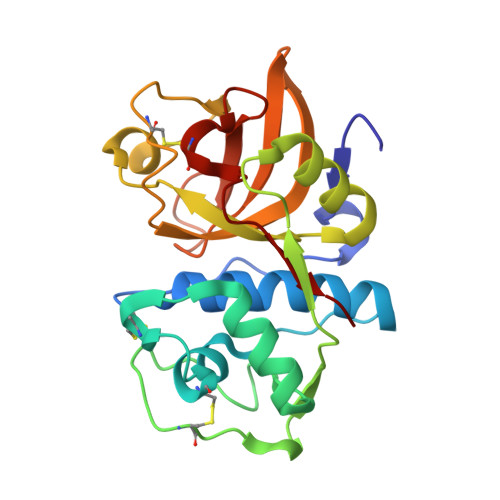
    Cathepsin K is a major drug target for osteoporosis and related-bone disorders. Using a combination of virtual combinatorial chemistry, QSAR modeling, and molecular docking studies, a series of cathepsin K inhibitors based on N-(functionalized benzoyl)-homocycloleucyl-glycinonitrile scaffold was developed. In order to avoid previous problems of cathepsin K inhibitors associated with lysosomotropism of compounds with basic character that resulted in off-target effects, a weakly- to nonbasic moiety was incorporated into the P3 position. Compounds 5, 6, and 9 were highly selective for cathepsin K when compared with cathepsins L and S, with the Ki values in the 10-30 nM range. The kinetic studies revealed that the new compounds exhibited reversible tight binding to cathepsin K, while the X-ray structural studies showed covalent and noncovalent binding between the nitrile group and the catalytic cysteine (Cys25) site.


  • Organizational Affiliation
    • National Institute of Chemistry, Hajdrihova 19, SI-1001 Ljubljana, Slovenia.

Macromolecule Content 

  • Total Structure Weight: 24.38 kDa 
  • Atom Count: 1,909 
  • Modeled Residue Count: 215 
  • Deposited Residue Count: 215 
  • Unique protein chains: 1

Macromolecules

Find similar proteins by:|  3D Structure
Entity ID: 1
MoleculeChains  Sequence LengthOrganismDetailsImage
Cathepsin K215Homo sapiensMutation(s): 0 
Gene Names: CTSKCTSOCTSO2
EC: 3.4.22.38
UniProt & NIH Common Fund Data Resources
Find proteins for P43235 (Homo sapiens)
Explore P43235 
Go to UniProtKB:  P43235
PHAROS:  P43235
GTEx:  ENSG00000143387 
Entity Groups
Sequence Clusters30% Identity50% Identity70% Identity90% Identity95% Identity100% Identity
UniProt GroupP43235
Sequence Annotations
Expand
Reference Sequence

Experimental Data & Validation

Experimental Data

  • Method: X-RAY DIFFRACTION
  • Resolution: 1.87 Å
  • R-Value Free:  0.237 (Depositor), 0.241 (DCC) 
  • R-Value Work:  0.181 (Depositor), 0.191 (DCC) 
  • R-Value Observed: 0.184 (Depositor) 
Space Group: P 21 21 21
Unit Cell:
Length ( Å )Angle ( ˚ )
a = 36.459α = 90
b = 54.45β = 90
c = 87.87γ = 90
Software Package:
Software NamePurpose
REFMACrefinement
SCALEPACKdata scaling
PHASERphasing
PDB_EXTRACTdata extraction
PROTEUM PLUSdata reduction

Structure Validation

View Full Validation Report


Ligand Structure Quality Assessment 


Entry History 

Deposition Data

Revision History  (Full details and data files)

  • Version 1.0: 2015-09-30
    Type: Initial release
  • Version 1.1: 2024-01-10
    Changes: Data collection, Database references, Derived calculations, Refinement description
  • Version 1.2: 2024-11-06
    Changes: Structure summary