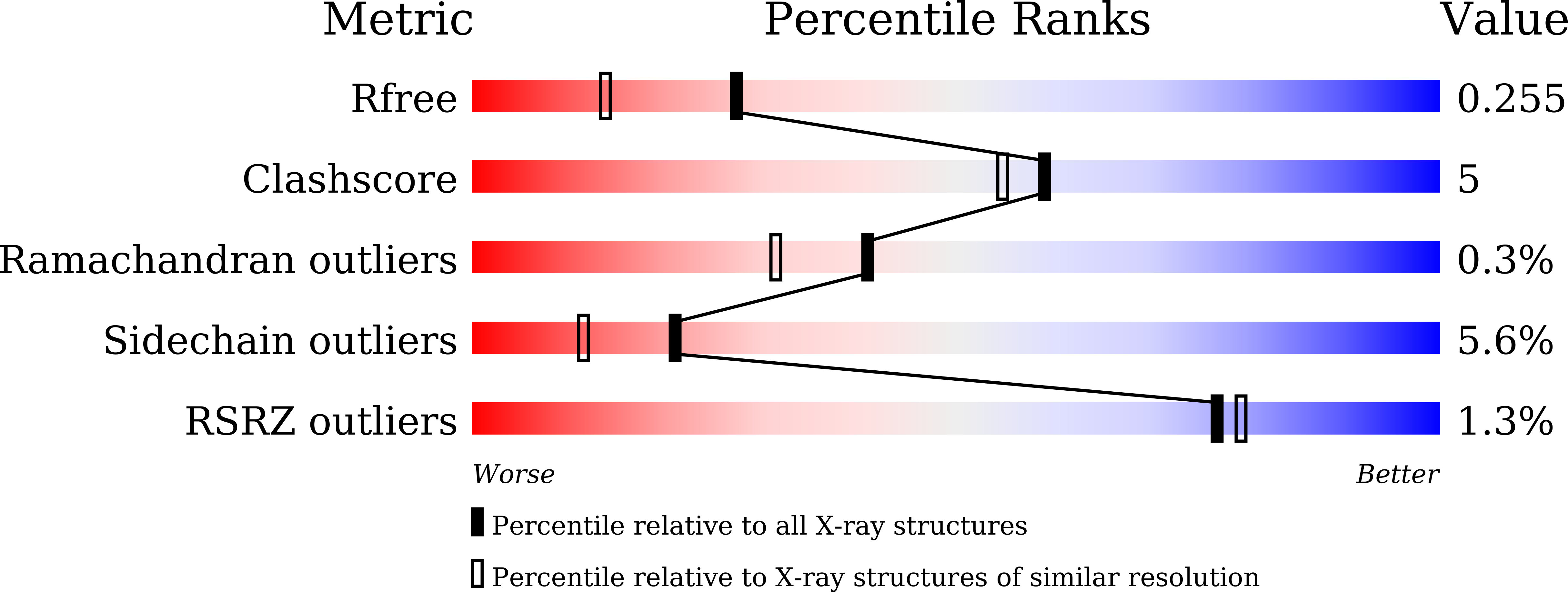
Structure of the 34 kDa F-actin-bundling protein ABP34 from Dictyostelium discoideum.
Kim, M.K., Kim, J.H., Kim, J.S., Kang, S.O.(2015) Acta Crystallogr D Biol Crystallogr 71: 1835-1849
- PubMed: 26327373
- DOI: https://doi.org/10.1107/S139900471501264X
- Primary Citation of Related Structures:
4X3N - PubMed Abstract:
The crystal structure of the 34 kDa F-actin-bundling protein ABP34 from Dictyostelium discoideum was solved by Ca(2+)/S-SAD phasing and refined at 1.89 Å resolution. ABP34 is a calcium-regulated actin-binding protein that cross-links actin filaments into bundles. Its in vitro F-actin-binding and F-actin-bundling activities were confirmed by a co-sedimentation assay and transmission electron microscopy. The co-localization of ABP34 with actin in cells was also verified. ABP34 adopts a two-domain structure with an EF-hand-containing N-domain and an actin-binding C-domain, but has no reported overall structural homologues. The EF-hand is occupied by a calcium ion with a pentagonal bipyramidal coordination as in the canonical EF-hand. The C-domain structure resembles a three-helical bundle and superposes well onto the rod-shaped helical structures of some cytoskeletal proteins. Residues 216-244 in the C-domain form part of the strongest actin-binding sites (193-254) and exhibit a conserved sequence with the actin-binding region of α-actinin and ABP120. Furthermore, the second helical region of the C-domain is kinked by a proline break, offering a convex surface towards the solvent area which is implicated in actin binding. The F-actin-binding model suggests that ABP34 binds to the side of the actin filament and residues 216-244 fit into a pocket between actin subdomains -1 and -2 through hydrophobic interactions. These studies provide insights into the calcium coordination in the EF-hand and F-actin-binding site in the C-domain of ABP34, which are associated through interdomain interactions.
Organizational Affiliation:
Laboratory of Biophysics, School of Biological Sciences, and Institute of Microbiology, Seoul National University, Seoul 151-742, Republic of Korea.


