4X28 | pdb_00004x28

Crystal structure of the ChsE4-ChsE5 complex from Mycobacterium tuberculosis


Experimental Data Snapshot

  • Method: X-RAY DIFFRACTION
  • Resolution: 1.99 Å
  • R-Value Free: 
    0.186 (Depositor), 0.195 (DCC) 
  • R-Value Work: 
    0.151 (Depositor), 0.161 (DCC) 
  • R-Value Observed: 
    0.153 (Depositor) 

wwPDB Validation 3D Report Full Report

Validation slider image for 4X28

Ligand Structure Quality Assessment 


This is version 1.4 of the entry. See complete history

Literature

Unraveling Cholesterol Catabolism in Mycobacterium tuberculosis: ChsE4-ChsE5 alpha 2 beta 2 Acyl-CoA Dehydrogenase Initiates beta-Oxidation of 3-Oxo-cholest-4-en-26-oyl CoA.

Yang, M.Lu, R.Guja, K.E.Wipperman, M.F.St Clair, J.R.Bonds, A.C.Garcia-Diaz, M.Sampson, N.S.

(2015) ACS Infect Dis 1: 110-125

  • DOI: https://doi.org/10.1021/id500033m
  • Primary Citation Related Structures: 
    4X28

  • PubMed Abstract: 

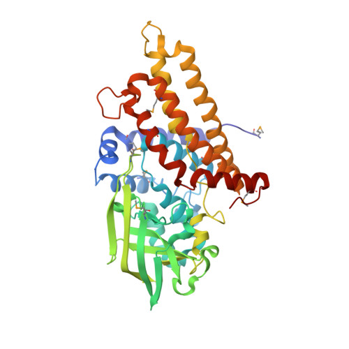
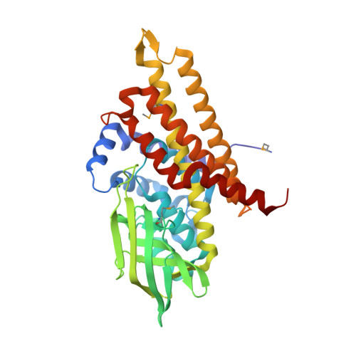
    The metabolism of host cholesterol by Mycobacterium tuberculosis ( Mtb ) is an important factor for both its virulence and pathogenesis, although how and why cholesterol metabolism is required is not fully understood. Mtb uses a unique set of catabolic enzymes that are homologous to those required for classical β-oxidation of fatty acids but are specific for steroid-derived substrates. Here, we identify and assign the substrate specificities of two of these enzymes, ChsE4-ChsE5 (Rv3504-Rv3505) and ChsE3 (Rv3573c), that carry out cholesterol side chain oxidation in Mtb. Steady-state assays demonstrate that ChsE4-ChsE5 preferentially catalyzes the oxidation of 3-oxo-cholest-4-en-26-oyl CoA in the first cycle of cholesterol side chain β-oxidation that ultimately yields propionyl-CoA, whereas ChsE3 specifically catalyzes the oxidation of 3-oxo-chol-4-en-24-oyl CoA in the second cycle of β-oxidation that generates acetyl-CoA. However, ChsE4-ChsE5 can catalyze the oxidation of 3-oxo-chol-4-en-24-oyl CoA as well as 3-oxo-4-pregnene-20-carboxyl-CoA. The functional redundancy of ChsE4-ChsE5 explains the in vivo phenotype of the igr knockout strain of Mycobacterium tuberculosis ; the loss of ChsE1-ChsE2 can be compensated for by ChsE4-ChsE5 during the chronic phase of infection. The X-ray crystallographic structure of ChsE4-ChsE5 was determined to a resolution of 2.0 Å and represents the first high-resolution structure of a heterotetrameric acyl-CoA dehydrogenase (ACAD). Unlike typical homotetrameric ACADs that bind four flavin adenine dinucleotide (FAD) cofactors, ChsE4-ChsE5 binds one FAD at each dimer interface, resulting in only two substrate-binding sites rather than the classical four active sites. A comparison of the ChsE4-ChsE5 substrate-binding site to those of known mammalian ACADs reveals an enlarged binding cavity that accommodates steroid substrates and highlights novel prospects for designing inhibitors against the committed β-oxidation step in the first cycle of cholesterol side chain degradation by Mtb .


  • Organizational Affiliation
    • Department of Chemistry, Department of Pharmacological Sciences, and Biochemistry and Structural Biology Graduate Program, Stony Brook University , Stony Brook, New York 11794, United States.

Macromolecule Content 

  • Total Structure Weight: 168.55 kDa 
  • Atom Count: 12,240 
  • Modeled Residue Count: 1,495 
  • Deposited Residue Count: 1,546 
  • Unique protein chains: 2

Macromolecules

Find similar proteins by:|  3D Structure
Entity ID: 1
MoleculeChains  Sequence LengthOrganismDetailsImage
Acyl-CoA dehydrogenase
A, B
400Mycobacterium tuberculosis H37RvMutation(s): 0 
Gene Names: fadE26Rv3504RVBD_3504LH57_19105P425_03646
EC: 1.3.99
UniProt
Find proteins for I6YCA3 (Mycobacterium tuberculosis (strain ATCC 25618 / H37Rv))
Explore I6YCA3 
Go to UniProtKB:  I6YCA3
Entity Groups
Sequence Clusters30% Identity50% Identity70% Identity90% Identity95% Identity100% Identity
UniProt GroupI6YCA3
Sequence Annotations
Expand
Reference Sequence
Find similar proteins by:|  3D Structure
Entity ID: 2
MoleculeChains  Sequence LengthOrganismDetailsImage
Acyl-CoA dehydrogenaseC [auth D],
D [auth C]
373Mycobacterium tuberculosis H37RvMutation(s): 0 
Gene Names: fadE27Rv3505RVBD_3505LH57_19110P425_03647
EC: 1.3.99
UniProt
Find proteins for I6Y3Q0 (Mycobacterium tuberculosis (strain ATCC 25618 / H37Rv))
Explore I6Y3Q0 
Go to UniProtKB:  I6Y3Q0
Entity Groups
Sequence Clusters30% Identity50% Identity70% Identity90% Identity95% Identity100% Identity
UniProt GroupI6Y3Q0
Sequence Annotations
Expand
Reference Sequence

Small Molecules

Modified Residues  1 Unique
IDChains TypeFormula2D DiagramParent
MSE
Query on MSE
A, B
L-PEPTIDE LINKINGC5 H11 N O2 SeMET

Experimental Data & Validation

Experimental Data

  • Method: X-RAY DIFFRACTION
  • Resolution: 1.99 Å
  • R-Value Free:  0.186 (Depositor), 0.195 (DCC) 
  • R-Value Work:  0.151 (Depositor), 0.161 (DCC) 
  • R-Value Observed: 0.153 (Depositor) 
Space Group: C 1 2 1
Unit Cell:
Length ( Å )Angle ( ˚ )
a = 185.745α = 90
b = 108.095β = 93.04
c = 82.019γ = 90
Software Package:
Software NamePurpose
REFMACrefinement
XDSdata reduction
Aimlessdata scaling
SHARPphasing

Structure Validation

View Full Validation Report


Ligand Structure Quality Assessment 


Entry History 

& Funding Information

Deposition Data


Funding OrganizationLocationGrant Number
National Institutes of Health/National Institute of General Medical Sciences (NIH/NIGMS)United StatesR01-GM100021
National Institutes of Health/National Institute of Environmental Health Sciences (NIH/NIEHS)United StatesF30-ES022930

Revision History  (Full details and data files)

  • Version 1.0: 2015-02-18
    Type: Initial release
  • Version 1.1: 2015-07-22
    Changes: Database references
  • Version 1.2: 2017-09-20
    Changes: Author supporting evidence, Derived calculations, Source and taxonomy, Structure summary
  • Version 1.3: 2019-12-18
    Changes: Author supporting evidence
  • Version 1.4: 2024-10-23
    Changes: Data collection, Database references, Refinement description, Structure summary