4WX9 | pdb_00004wx9

Crystal structure of Mycobacterium tuberculosis OGT in complex with DNA


Experimental Data Snapshot

  • Method: X-RAY DIFFRACTION
  • Resolution: 3.00 Å
  • R-Value Free: 
    0.265 (Depositor), 0.258 (DCC) 
  • R-Value Work: 
    0.194 (Depositor), 0.199 (DCC) 
  • R-Value Observed: 
    0.197 (Depositor) 

Starting Models: experimental
View more details

wwPDB Validation 3D Report Full Report

Validation slider image for 4WX9

This is version 1.3 of the entry. See complete history

Literature

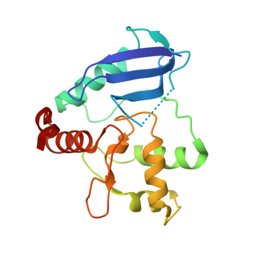
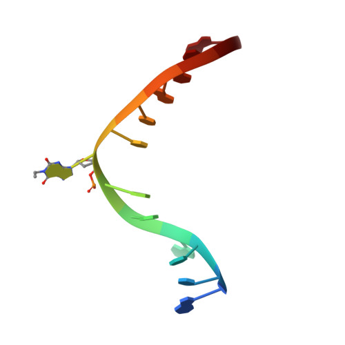
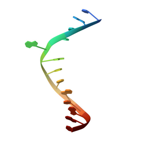
Crystal structure of Mycobacterium tuberculosis O6-methylguanine-DNA methyltransferase protein clusters assembled on to damaged DNA.

Miggiano, R.Perugino, G.Ciaramella, M.Serpe, M.Rejman, D.Pav, O.Pohl, R.Garavaglia, S.Lahiri, S.Rizzi, M.Rossi, F.

(2016) Biochem J 473: 123-133

  • DOI: https://doi.org/10.1042/BJ20150833
  • Primary Citation Related Structures: 
    4WX9, 4WXC, 4WXD

  • PubMed Abstract: 

    Mycobacterium tuberculosis O(6)-methylguanine-DNA methyltransferase (MtOGT) contributes to protect the bacterial GC-rich genome against the pro-mutagenic potential of O(6)-methylated guanine in DNA. Several strains of M. tuberculosis found worldwide encode a point-mutated O(6)-methylguanine-DNA methyltransferase (OGT) variant (MtOGT-R37L), which displays an arginine-to-leucine substitution at position 37 of the poorly functionally characterized N-terminal domain of the protein. Although the impact of this mutation on the MtOGT activity has not yet been proved in vivo, we previously demonstrated that a recombinant MtOGT-R37L variant performs a suboptimal alkylated-DNA repair in vitro, suggesting a direct role for the Arg(37)-bearing region in catalysis. The crystal structure of MtOGT complexed with modified DNA solved in the present study reveals details of the protein-protein and protein-DNA interactions occurring during alkylated-DNA binding, and the protein capability also to host unmodified bases inside the active site, in a fully extrahelical conformation. Our data provide the first experimental picture at the atomic level of a possible mode of assembling three adjacent MtOGT monomers on the same monoalkylated dsDNA molecule, and disclose the conformational flexibility of discrete regions of MtOGT, including the Arg(37)-bearing random coil. This peculiar structural plasticity of MtOGT could be instrumental to proper protein clustering at damaged DNA sites, as well as to protein-DNA complexes disassembling on repair.


  • Organizational Affiliation
    • DSF-Dipartimento di Scienze del Farmaco, University of Piemonte Orientale, 28100 Novara, Italy.

Macromolecule Content 

  • Total Structure Weight: 61.66 kDa 
  • Atom Count: 4,271 
  • Modeled Residue Count: 515 
  • Deposited Residue Count: 521 
  • Unique protein chains: 1
  • Unique nucleic acid chains: 2

Macromolecules


Find similar proteins by:|  3D Structure
Entity ID: 3
MoleculeChains  Sequence LengthOrganismDetailsImage
Methylated-DNA--protein-cysteine methyltransferaseC [auth A],
D [auth B],
E [auth C]
165Mycobacterium tuberculosis H37RvMutation(s): 1 
Gene Names: ogtRv1316cMTCY130.01c
EC: 2.1.1.63
UniProt
Find proteins for P9WJW5 (Mycobacterium tuberculosis (strain ATCC 25618 / H37Rv))
Explore P9WJW5 
Go to UniProtKB:  P9WJW5
Entity Groups
Sequence Clusters30% Identity50% Identity70% Identity90% Identity95% Identity100% Identity
UniProt GroupP9WJW5
Sequence Annotations
Expand
Reference Sequence
Find similar nucleic acids by:  Sequence
Entity ID: 1
MoleculeChains LengthOrganismImage
DNA (5'-D(*GP*CP*CP*AP*TP*GP*(E1X)P*CP*TP*AP*GP*TP*A)-3')A [auth D]13synthetic construct
Sequence Annotations
Expand
Reference Sequence
Find similar nucleic acids by:  Sequence
Entity ID: 2
MoleculeChains LengthOrganismImage
DNA (5'-D(P*TP*AP*CP*TP*AP*GP*CP*CP*AP*TP*GP*GP*C)-3')B [auth E]13synthetic construct
Sequence Annotations
Expand
Reference Sequence

Small Molecules

Ligands 1 Unique
IDChains Name / Formula / InChI Key2D Diagram3D Interactions
GOL

Query on GOL


Download:Ideal Coordinates CCD File
F [auth A]GLYCEROL
C3 H8 O3
PEDCQBHIVMGVHV-UHFFFAOYSA-N

Experimental Data & Validation

Experimental Data

  • Method: X-RAY DIFFRACTION
  • Resolution: 3.00 Å
  • R-Value Free:  0.265 (Depositor), 0.258 (DCC) 
  • R-Value Work:  0.194 (Depositor), 0.199 (DCC) 
  • R-Value Observed: 0.197 (Depositor) 
Space Group: P 21 21 21
Unit Cell:
Length ( Å )Angle ( ˚ )
a = 43.481α = 90
b = 102.905β = 90
c = 137.093γ = 90
Software Package:
Software NamePurpose
REFMACrefinement
XDSdata reduction
SCALAdata scaling
PHASERphasing

Structure Validation

View Full Validation Report


Entry History 

& Funding Information

Deposition Data


Funding OrganizationLocationGrant Number
European CommunityItalySysteMTB HEALTH-F4-2010-241587

Revision History  (Full details and data files)

  • Version 1.0: 2015-11-04
    Type: Initial release
  • Version 1.1: 2015-11-11
    Changes: Database references
  • Version 1.2: 2016-01-13
    Changes: Database references
  • Version 1.3: 2024-01-10
    Changes: Data collection, Database references, Refinement description