


Structure of the PTP-like phytase from Selenomonas ruminantium in complex with myo-inositol-(1,4,5)-trikisphosphate
Bruder, L.M.To be published.
Experimental Data Snapshot
Starting Model: experimental
View more details
Entity ID: 1 | |||||
|---|---|---|---|---|---|
| Molecule | Chains | Sequence Length | Organism | Details | Image |
| Myo-inositol phosphohydrolase | 342 | Pseudoselenomonas ruminantium | Mutation(s): 1 Gene Names: phyA EC: 3.1.3.8 | 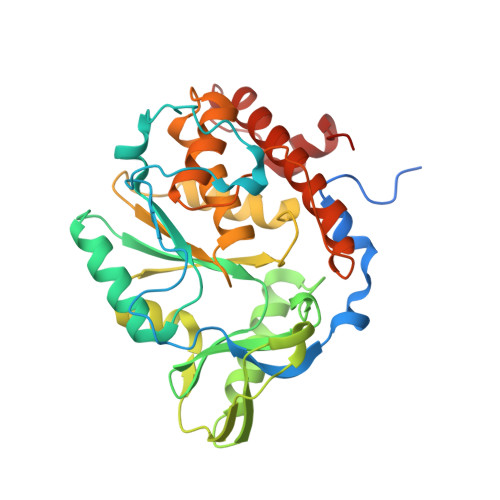 | |
UniProt | |||||
Entity Groups | |||||
| Sequence Clusters | 30% Identity50% Identity70% Identity90% Identity95% Identity100% Identity | ||||
| UniProt Group | Q7WUJ1 | ||||
Sequence AnnotationsExpand | |||||
Reference Sequence | |||||
| Ligands 4 Unique | |||||
|---|---|---|---|---|---|
| ID | Chains | Name / Formula / InChI Key | 2D Diagram | 3D Interactions | |
| I3P Download:Ideal Coordinates CCD File | C [auth A], G [auth B] | D-MYO-INOSITOL-1,4,5-TRIPHOSPHATE C6 H15 O15 P3 MMWCIQZXVOZEGG-XJTPDSDZSA-N |  | ||
| PO4 Download:Ideal Coordinates CCD File | D [auth A], H [auth B] | PHOSPHATE ION O4 P NBIIXXVUZAFLBC-UHFFFAOYSA-K |  | ||
| GOL Download:Ideal Coordinates CCD File | E [auth A], F [auth A], J [auth B], K [auth B] | GLYCEROL C3 H8 O3 PEDCQBHIVMGVHV-UHFFFAOYSA-N |  | ||
| CL Download:Ideal Coordinates CCD File | I [auth B] | CHLORIDE ION Cl VEXZGXHMUGYJMC-UHFFFAOYSA-M |  | ||
| Length ( Å ) | Angle ( ˚ ) |
|---|---|
| a = 45.98 | α = 90 |
| b = 137.67 | β = 102.42 |
| c = 80.01 | γ = 90 |
| Software Name | Purpose |
|---|---|
| REFMAC | refinement |
| MOSFLM | data reduction |
| Aimless | data scaling |
| Funding Organization | Location | Grant Number |
|---|---|---|
| Canada Foundation for Innovation | Canada | -- |
| Natural Sciences and Engineering Research Council (NSERC, Canada) | Canada | -- |


