4WNX | pdb_00004wnx

Netrin 4 lacking the C-terminal Domain


Experimental Data Snapshot

  • Method: X-RAY DIFFRACTION
  • Resolution: 2.72 Å
  • R-Value Free: 
    0.263 (Depositor), 0.268 (DCC) 
  • R-Value Work: 
    0.220 (Depositor), 0.228 (DCC) 
  • R-Value Observed: 
    0.222 (Depositor) 

Starting Model: experimental
View more details

wwPDB Validation 3D Report Full Report

Validation slider image for 4WNX

Ligand Structure Quality Assessment 


This is version 2.2 of the entry. See complete history

Literature

Structural decoding of netrin-4 reveals a regulatory function towards mature basement membranes.

Reuten, R.Patel, T.R.McDougall, M.Rama, N.Nikodemus, D.Gibert, B.Delcros, J.G.Prein, C.Meier, M.Metzger, S.Zhou, Z.Kaltenberg, J.McKee, K.K.Bald, T.Tuting, T.Zigrino, P.Djonov, V.Bloch, W.Clausen-Schaumann, H.Poschl, E.Yurchenco, P.D.Ehrbar, M.Mehlen, P.Stetefeld, J.Koch, M.

(2016) Nat Commun 7: 13515-13515

  • DOI: https://doi.org/10.1038/ncomms13515
  • Primary Citation Related Structures: 
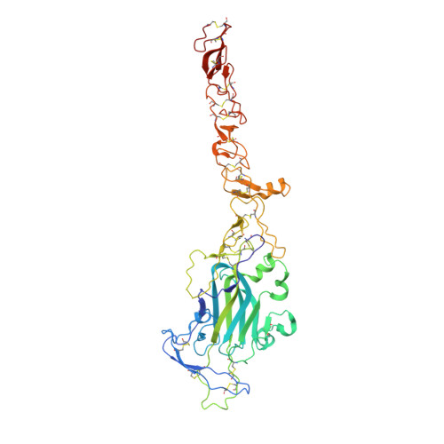
    4WNX

  • PubMed Abstract: 

    Netrins, a family of laminin-related molecules, have been proposed to act as guidance cues either during nervous system development or the establishment of the vascular system. This was clearly demonstrated for netrin-1 via its interaction with the receptors DCC and UNC5s. However, mainly based on shared homologies with netrin-1, netrin-4 was also proposed to play a role in neuronal outgrowth and developmental/pathological angiogenesis via interactions with netrin-1 receptors. Here, we present the high-resolution structure of netrin-4, which shows unique features in comparison with netrin-1, and show that it does not bind directly to any of the known netrin-1 receptors. We show that netrin-4 disrupts laminin networks and basement membranes (BMs) through high-affinity binding to the laminin γ1 chain. We hypothesize that this laminin-related function is essential for the previously described effects on axon growth promotion and angiogenesis. Our study unveils netrin-4 as a non-enzymatic extracellular matrix protein actively disrupting pre-existing BMs.


  • Organizational Affiliation
    • Institute for Dental Research and Oral Musculoskeletal Biology, Medical Faculty, University of Cologne, Joseph-Stelzmann-Strasse 52, Cologne 50931, Germany.

Macromolecule Content 

  • Total Structure Weight: 49.64 kDa 
  • Atom Count: 3,382 
  • Modeled Residue Count: 429 
  • Deposited Residue Count: 433 
  • Unique protein chains: 1

Macromolecules

Find similar proteins by:|  3D Structure
Entity ID: 1
MoleculeChains  Sequence LengthOrganismDetailsImage
Netrin-4433Mus musculusMutation(s): 0 
Gene Names: Ntn4
UniProt & NIH Common Fund Data Resources
Find proteins for Q9JI33 (Mus musculus)
Explore Q9JI33 
Go to UniProtKB:  Q9JI33
IMPC:  MGI:1888978
Entity Groups
Sequence Clusters30% Identity50% Identity70% Identity90% Identity95% Identity100% Identity
UniProt GroupQ9JI33
Glycosylation
Glycosylation Sites: 3Go to GlyGen: Q9JI33-1
Sequence Annotations
Expand
Reference Sequence

Oligosaccharides

Help  
Entity ID: 2
MoleculeChains Length2D Diagram GlycosylationD Interactions
2-acetamido-2-deoxy-beta-D-glucopyranose-(1-4)-2-acetamido-2-deoxy-beta-D-glucopyranose
B, C
2N-Glycosylation
Glycosylation Resources
GlyTouCan: G42666HT
GlyCosmos: G42666HT
GlyGen: G42666HT

Experimental Data & Validation

Experimental Data

  • Method: X-RAY DIFFRACTION
  • Resolution: 2.72 Å
  • R-Value Free:  0.263 (Depositor), 0.268 (DCC) 
  • R-Value Work:  0.220 (Depositor), 0.228 (DCC) 
  • R-Value Observed: 0.222 (Depositor) 
Space Group: C 1 2 1
Unit Cell:
Length ( Å )Angle ( ˚ )
a = 107.755α = 90
b = 75.049β = 96.07
c = 74.672γ = 90
Software Package:
Software NamePurpose
CrystalCleardata collection
HKL-2000data scaling
SCALEPACKdata scaling
PDB_EXTRACTdata extraction
PHENIXrefinement
Cootmodel building
PHASERphasing
REFMACrefinement
DENZOdata reduction

Structure Validation

View Full Validation Report


Ligand Structure Quality Assessment 


Entry History 

Deposition Data

Revision History  (Full details and data files)

  • Version 1.0: 2016-02-10
    Type: Initial release
  • Version 1.1: 2016-12-14
    Changes: Database references
  • Version 1.2: 2017-11-22
    Changes: Refinement description
  • Version 2.0: 2020-07-29
    Type: Remediation
    Reason: Carbohydrate remediation
    Changes: Atomic model, Data collection, Derived calculations, Structure summary
  • Version 2.1: 2023-09-27
    Changes: Data collection, Database references, Refinement description, Structure summary
  • Version 2.2: 2024-10-16
    Changes: Structure summary