4WMZ | pdb_00004wmz

S. cerevisiae CYP51 complexed with fluconazole in the active site


Experimental Data Snapshot

  • Method: X-RAY DIFFRACTION
  • Resolution: 2.05 Å
  • R-Value Free: 
    0.232 (Depositor), 0.233 (DCC) 
  • R-Value Work: 
    0.199 (Depositor), 0.203 (DCC) 
  • R-Value Observed: 
    0.200 (Depositor) 

Starting Model: experimental
View more details

wwPDB Validation 3D Report Full Report

Validation slider image for 4WMZ

Ligand Structure Quality Assessment 


This is version 1.4 of the entry. See complete history

Literature

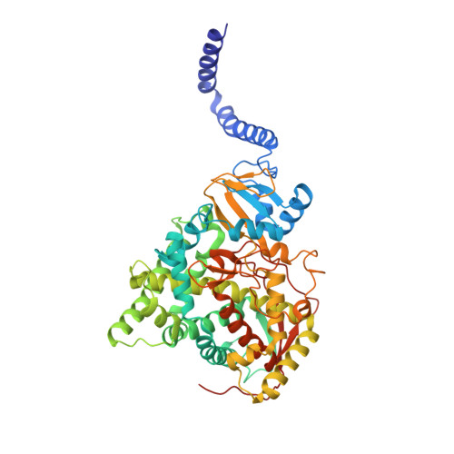
Structural Insights into Binding of the Antifungal Drug Fluconazole to Saccharomyces cerevisiae Lanosterol 14 alpha-Demethylase.

Sagatova, A.A.Keniya, M.V.Wilson, R.K.Monk, B.C.Tyndall, J.D.

(2015) Antimicrob Agents Chemother 59: 4982-4989

  • DOI: https://doi.org/10.1128/AAC.00925-15
  • Primary Citation Related Structures: 
    4WMZ

  • PubMed Abstract: 

    Infections by fungal pathogens such as Candida albicans and Aspergillus fumigatus and their resistance to triazole drugs are major concerns. Fungal lanosterol 14α-demethylase belongs to the CYP51 class in the cytochrome P450 superfamily of enzymes. This monospanning bitopic membrane protein is involved in ergosterol biosynthesis and is the primary target of azole antifungal drugs, including fluconazole. The lack of high-resolution structural information for this drug target from fungal pathogens has been a limiting factor for the design of modified triazole drugs that will overcome resistance. Here we report the X-ray structure of full-length Saccharomyces cerevisiae lanosterol 14α-demethylase in complex with fluconazole at a resolution of 2.05 Å. This structure shows the key interactions involved in fluconazole binding and provides insight into resistance mechanisms by revealing a water-mediated hydrogen bonding network between the drug and tyrosine 140, a residue frequently found mutated to histidine or phenylalanine in resistant clinical isolates.


  • Organizational Affiliation
    • Sir John Walsh Research Institute, University of Otago, Dunedin, New Zealand.

Macromolecule Content 

  • Total Structure Weight: 62.81 kDa 
  • Atom Count: 4,481 
  • Modeled Residue Count: 527 
  • Deposited Residue Count: 539 
  • Unique protein chains: 1

Macromolecules

Find similar proteins by:|  3D Structure
Entity ID: 1
MoleculeChains  Sequence LengthOrganismDetailsImage
Lanosterol 14-alpha demethylase539Saccharomyces cerevisiae YJM789Mutation(s): 0 
Gene Names: ERG11SCY_2394
EC: 1.14.14.154
Membrane Entity: Yes 
UniProt
Find proteins for A6ZSR0 (Saccharomyces cerevisiae (strain YJM789))
Explore A6ZSR0 
Go to UniProtKB:  A6ZSR0
Entity Groups
Sequence Clusters30% Identity50% Identity70% Identity90% Identity95% Identity100% Identity
UniProt GroupA6ZSR0
Sequence Annotations
Expand
Reference Sequence

Small Molecules

Ligands 2 Unique
IDChains Name / Formula / InChI Key2D Diagram3D Interactions
HEM

Query on HEM


Download:Ideal Coordinates CCD File
B [auth A]PROTOPORPHYRIN IX CONTAINING FE
C34 H32 Fe N4 O4
KABFMIBPWCXCRK-RGGAHWMASA-L
TPF

Query on TPF


Download:Ideal Coordinates CCD File
C [auth A]2-(2,4-DIFLUOROPHENYL)-1,3-DI(1H-1,2,4-TRIAZOL-1-YL)PROPAN-2-OL
C13 H12 F2 N6 O
RFHAOTPXVQNOHP-UHFFFAOYSA-N

Experimental Data & Validation

Experimental Data

  • Method: X-RAY DIFFRACTION
  • Resolution: 2.05 Å
  • R-Value Free:  0.232 (Depositor), 0.233 (DCC) 
  • R-Value Work:  0.199 (Depositor), 0.203 (DCC) 
  • R-Value Observed: 0.200 (Depositor) 
Space Group: P 1 21 1
Unit Cell:
Length ( Å )Angle ( ˚ )
a = 77.24α = 90
b = 65.21β = 98.43
c = 81.02γ = 90
Software Package:
Software NamePurpose
PHENIXrefinement
Aimlessdata scaling

Structure Validation

View Full Validation Report


Ligand Structure Quality Assessment 


Entry History 

& Funding Information

Deposition Data


Funding OrganizationLocationGrant Number
Health Research Council (HRC)New Zealand--
Marsden FundNew Zealand--

Revision History  (Full details and data files)

  • Version 1.0: 2015-07-01
    Type: Initial release
  • Version 1.1: 2015-07-29
    Changes: Database references
  • Version 1.2: 2017-09-20
    Changes: Data collection, Database references, Derived calculations, Source and taxonomy
  • Version 1.3: 2020-01-01
    Changes: Author supporting evidence
  • Version 1.4: 2023-09-27
    Changes: Data collection, Database references, Refinement description