4WMR | pdb_00004wmr

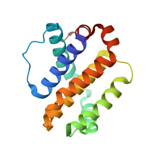
STRUCTURE OF MCL1 BOUND TO BRD inhibitor ligand 1 AT 1.7A


Experimental Data Snapshot

  • Method: X-RAY DIFFRACTION
  • Resolution: 1.70 Å
  • R-Value Free: 
    0.206 (Depositor), 0.215 (DCC) 
  • R-Value Work: 
    0.169 (Depositor), 0.179 (DCC) 
  • R-Value Observed: 
    0.171 (Depositor) 

wwPDB Validation 3D Report Full Report

Validation slider image for 4WMR

Ligand Structure Quality Assessment 


This is version 1.1 of the entry. See complete history. 

Literature

A Maltose-Binding Protein Fusion Construct Yields a Robust Crystallography Platform for MCL1.

Clifton, M.C., Dranow, D.M., Leed, A., Fulroth, B., Fairman, J.W., Abendroth, J., Atkins, K.A., Wallace, E., Fan, D., Xu, G., Ni, Z.J., Daniels, D., Van Drie, J., Wei, G., Burgin, A.B., Golub, T.R., Hubbard, B.K., Serrano-Wu, M.H.

(2015) PLoS One 10: e0125010-e0125010

  • DOI: https://doi.org/10.1371/journal.pone.0125010
  • Primary Citation Related Structures: 
    4WMR, 4WMS, 4WMT, 4WMU, 4WMV, 4WMW, 4WMX

  • PubMed Abstract: 

    Crystallization of a maltose-binding protein MCL1 fusion has yielded a robust crystallography platform that generated the first apo MCL1 crystal structure, as well as five ligand-bound structures. The ability to obtain fragment-bound structures advances structure-based drug design efforts that, despite considerable effort, had previously been intractable by crystallography. In the ligand-independent crystal form we identify inhibitor binding modes not observed in earlier crystallographic systems. This MBP-MCL1 construct dramatically improves the structural understanding of well-validated MCL1 ligands, and will likely catalyze the structure-based optimization of high affinity MCL1 inhibitors.


  • Organizational Affiliation: 
    • Beryllium, Bedford, Massachusetts, United States of America.

Macromolecule Content 

  • Total Structure Weight: 18.34 kDa 
  • Atom Count: 1,373 
  • Modeled Residue Count: 150 
  • Deposited Residue Count: 150 
  • Unique protein chains: 1

Macromolecules

Find similar proteins by:|  3D Structure
Entity ID: 1
MoleculeChains  Sequence LengthOrganismDetailsImage
Induced myeloid leukemia cell differentiation protein Mcl-1150Homo sapiensMutation(s): 0 
Gene Names: MCL1, BCL2L3
UniProt & NIH Common Fund Data Resources
Find proteins for Q07820 (Homo sapiens)
Explore Q07820 
Go to UniProtKB:  Q07820
PHAROS:  Q07820
GTEx:  ENSG00000143384 
Entity Groups
Sequence Clusters30% Identity50% Identity70% Identity90% Identity95% Identity100% Identity
UniProt GroupQ07820
Sequence Annotations
Expand
Reference Sequence

Small Molecules

Ligands 3 Unique
IDChains Name / Formula / InChI Key2D Diagram3D Interactions
865

Query on 865


Download:Ideal Coordinates CCD File
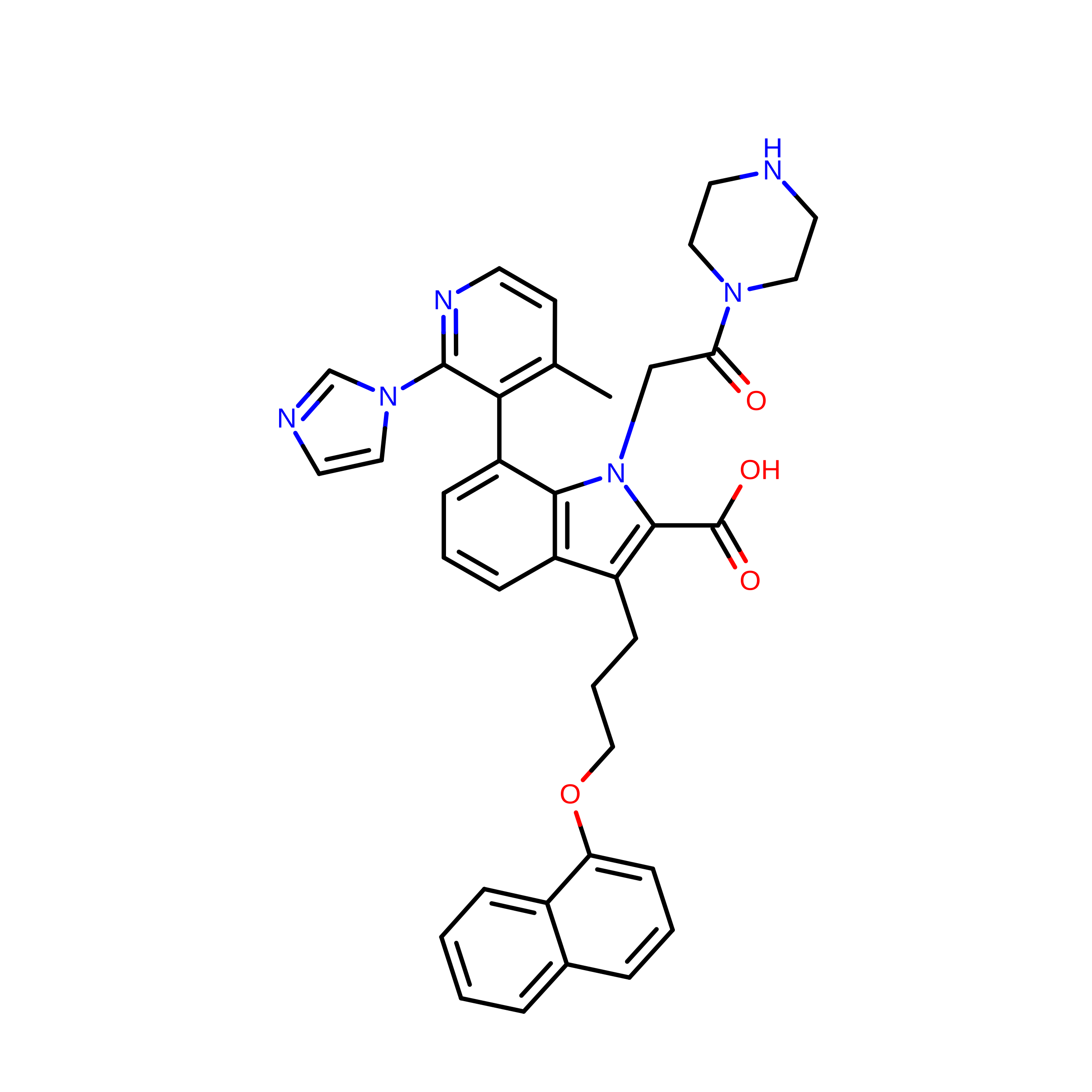
E [auth A]7-[2-(1H-imidazol-1-yl)-4-methylpyridin-3-yl]-3-[3-(naphthalen-1-yloxy)propyl]-1-[2-oxo-2-(piperazin-1-yl)ethyl]-1H-indole-2-carboxylic acid
C37 H36 N6 O4
ZZKICOFWGKKSRR-UHFFFAOYSA-N
POP

Query on POP


Download:Ideal Coordinates CCD File
F [auth A],
G [auth A]
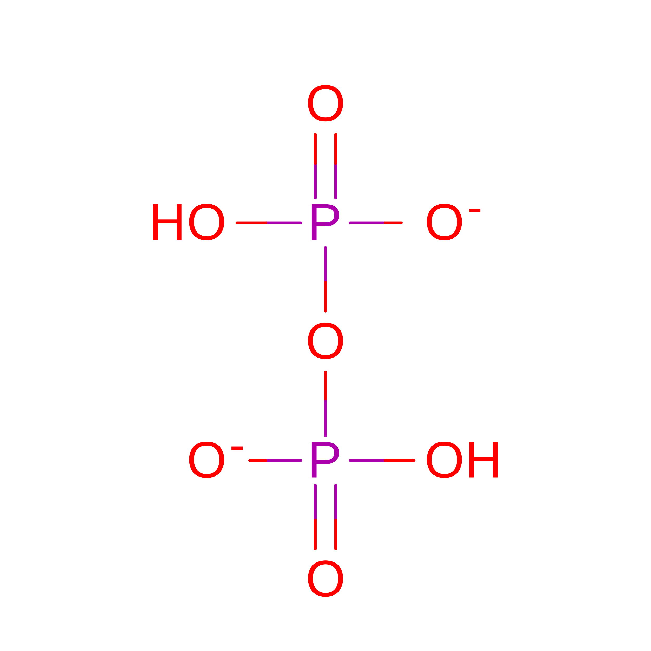
PYROPHOSPHATE 2-
H2 O7 P2
XPPKVPWEQAFLFU-UHFFFAOYSA-L
ZN

Query on ZN


Download:Ideal Coordinates CCD File
B [auth A],
C [auth A],
D [auth A]
ZINC ION
Zn
PTFCDOFLOPIGGS-UHFFFAOYSA-N

Experimental Data & Validation

Experimental Data

  • Method: X-RAY DIFFRACTION
  • Resolution: 1.70 Å
  • R-Value Free:  0.206 (Depositor), 0.215 (DCC) 
  • R-Value Work:  0.169 (Depositor), 0.179 (DCC) 
  • R-Value Observed: 0.171 (Depositor) 
Space Group: C 1 2 1
Unit Cell:
Length ( Å )Angle ( ˚ )
a = 72.77α = 90
b = 38.41β = 102.6
c = 48.33γ = 90
Software Package:
Software NamePurpose
XDSdata reduction
REFMACrefinement
PDB_EXTRACTdata extraction

Structure Validation

View Full Validation Report


Ligand Structure Quality Assessment 


Entry History 

Deposition Data

Revision History  (Full details and data files)

  • Version 1.0: 2015-05-06
    Type: Initial release
  • Version 1.1: 2023-12-27
    Changes: Data collection, Database references, Derived calculations, Other, Refinement description, Source and taxonomy, Structure summary