


Crystal structure of oxaloacetate and NADH bound MDH2
Eo, Y.M., Han, B.G., Ahn, H.C.To be published.
Experimental Data Snapshot
Entity ID: 1 | |||||
|---|---|---|---|---|---|
| Molecule | Chains | Sequence Length | Organism | Details | Image |
| Malate dehydrogenase, mitochondrial | 340 | Homo sapiens | Mutation(s): 0 Gene Names: MDH2 EC: 1.1.1.37 | 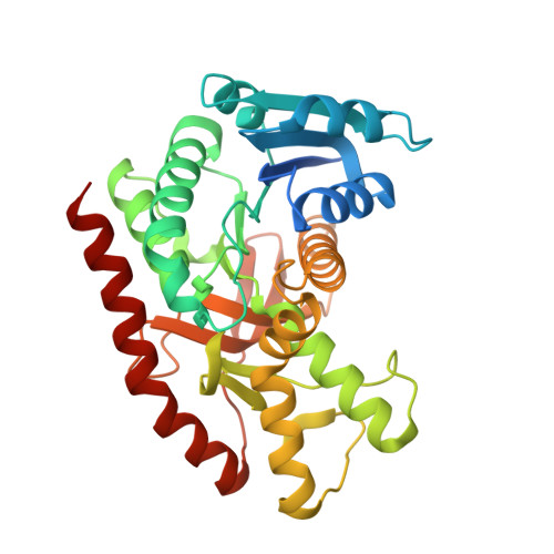 | |
UniProt & NIH Common Fund Data Resources | |||||
PHAROS: P40926 GTEx: ENSG00000146701 | |||||
Entity Groups | |||||
| Sequence Clusters | 30% Identity50% Identity70% Identity90% Identity95% Identity100% Identity | ||||
| UniProt Group | P40926 | ||||
Sequence AnnotationsExpand | |||||
Reference Sequence | |||||
| Ligands 2 Unique | |||||
|---|---|---|---|---|---|
| ID | Chains | Name / Formula / InChI Key | 2D Diagram | 3D Interactions | |
| NAI Download:Ideal Coordinates CCD File | E [auth A], G [auth B], I [auth C], K [auth D] | 1,4-DIHYDRONICOTINAMIDE ADENINE DINUCLEOTIDE C21 H29 N7 O14 P2 BOPGDPNILDQYTO-NNYOXOHSSA-N |  | ||
| OAA Download:Ideal Coordinates CCD File | F [auth A], H [auth B], J [auth C], L [auth D] | OXALOACETATE ION C4 H3 O5 KHPXUQMNIQBQEV-UHFFFAOYSA-M |  | ||
| Length ( Å ) | Angle ( ˚ ) |
|---|---|
| a = 59.989 | α = 90 |
| b = 152.191 | β = 90 |
| c = 155.869 | γ = 90 |
| Software Name | Purpose |
|---|---|
| REFMAC | refinement |


