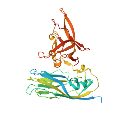
4WIZ | pdb_00004wiz

Crystal structure of Grouper nervous necrosis virus-like particle at 3.6A


Experimental Data Snapshot

  • Method: X-RAY DIFFRACTION
  • Resolution: 3.60 Å
  • R-Value Free: 
    0.297 (Depositor), 0.294 (DCC) 
  • R-Value Work: 
    0.255 (Depositor), 0.255 (DCC) 
  • R-Value Observed: 
    0.257 (Depositor) 

wwPDB Validation 3D Report Full Report

Validation slider image for 4WIZ

This is version 1.4 of the entry. See complete history. 

Literature

Crystal Structures of a Piscine Betanodavirus: Mechanisms of Capsid Assembly and Viral Infection

Chen, N.C., Yoshimura, M., Guan, H.H., Wang, T.Y., Misumi, Y., Lin, C.C., Chuankhayan, P., Nakagawa, A., Chan, S.I., Tsukihara, T., Chen, T.Y., Chen, C.J.

(2015) PLoS Pathog 11: e1005203-e1005203

  • DOI: https://doi.org/10.1371/journal.ppat.1005203
  • Primary Citation Related Structures: 
    4RFT, 4RFU, 4WIZ

  • PubMed Abstract: 

    Betanodaviruses cause massive mortality in marine fish species with viral nervous necrosis. The structure of a T = 3 Grouper nervous necrosis virus-like particle (GNNV-LP) is determined by the ab initio method with non-crystallographic symmetry averaging at 3.6 Å resolution. Each capsid protein (CP) shows three major domains: (i) the N-terminal arm, an inter-subunit extension at the inner surface; (ii) the shell domain (S-domain), a jelly-roll structure; and (iii) the protrusion domain (P-domain) formed by three-fold trimeric protrusions. In addition, we have determined structures of the T = 1 subviral particles (SVPs) of (i) the delta-P-domain mutant (residues 35-217) at 3.1 Å resolution; and (ii) the N-ARM deletion mutant (residues 35-338) at 7 Å resolution; and (iii) the structure of the individual P-domain (residues 214-338) at 1.2 Å resolution. The P-domain reveals a novel DxD motif asymmetrically coordinating two Ca2+ ions, and seems to play a prominent role in the calcium-mediated trimerization of the GNNV CPs during the initial capsid assembly process. The flexible N-ARM (N-terminal arginine-rich motif) appears to serve as a molecular switch for T = 1 or T = 3 assembly. Finally, we find that polyethylene glycol, which is incorporated into the P-domain during the crystallization process, enhances GNNV infection. The present structural studies together with the biological assays enhance our understanding of the role of the P-domain of GNNV in the capsid assembly and viral infection by this betanodavirus.


  • Organizational Affiliation: 
    • Institute of Biotechnology, National Cheng Kung University, Tainan, Taiwan; Life Science Group, Scientific Research Division, National Synchrotron Radiation Research Center, Hsinchu, Taiwan.

Macromolecule Content 

  • Total Structure Weight: 3,344.02 kDa 
  • Atom Count: 203,250 
  • Modeled Residue Count: 26,310 
  • Deposited Residue Count: 30,420 
  • Unique protein chains: 1

Macromolecules

Find similar proteins by:|  3D Structure
Entity ID: 1
MoleculeChains  Sequence LengthOrganismDetailsImage
Coat protein338Epinephelus coioides nervous necrosis virusMutation(s): 1 
UniProt
Find proteins for Q8JNX5 (Epinephelus coioides nervous necrosis virus)
Explore Q8JNX5 
Go to UniProtKB:  Q8JNX5
Entity Groups
Sequence Clusters30% Identity50% Identity70% Identity90% Identity95% Identity100% Identity
UniProt GroupQ8JNX5
Sequence Annotations
Expand
Reference Sequence

Small Molecules

Ligands 1 Unique
IDChains Name / Formula / InChI Key2D Diagram3D Interactions
CA

Query on CA


Download:Ideal Coordinates CCD File
AD [auth AE]
AE [auth BN]
AF [auth BW]
BD [auth BF]
BE [auth AN]
AD [auth AE],
AE [auth BN],
AF [auth BW],
BD [auth BF],
BE [auth AN],
BF [auth BW],
CD [auth BF],
CE [auth BO],
CF [auth AW],
DD [auth AF],
DE [auth BO],
DF [auth BX],
ED [auth BG],
EE [auth AO],
EF [auth BX],
FD [auth BG],
FE [auth BP],
FF [auth AX],
GD [auth AG],
GE [auth BP],
GF [auth BY],
HD [auth BH],
HE [auth AP],
HF [auth BY],
ID [auth BH],
IE [auth BQ],
IF [auth AY],
JD [auth AH],
JE [auth BQ],
JF [auth BZ],
KD [auth BI],
KE [auth AQ],
KF [auth BZ],
LD [auth BI],
LE [auth BR],
LF [auth AZ],
MC [auth BA],
MD [auth AI],
ME [auth BR],
MF [auth Ba],
NC [auth BA],
ND [auth BJ],
NE [auth AR],
NF [auth Ba],
OC [auth AA],
OD [auth BJ],
OE [auth BS],
OF [auth Aa],
PC [auth BB],
PD [auth AJ],
PE [auth BS],
PF [auth Bb],
QC [auth BB],
QD [auth BK],
QE [auth AS],
QF [auth Bb],
RC [auth AB],
RD [auth BK],
RE [auth BT],
RF [auth Ab],
SC [auth BC],
SD [auth AK],
SE [auth BT],
SF [auth Bc],
TC [auth BC],
TD [auth BL],
TE [auth AT],
TF [auth Bc],
UC [auth AC],
UD [auth BL],
UE [auth BU],
UF [auth Ac],
VC [auth BD],
VD [auth AL],
VE [auth BU],
VF [auth Bd],
WC [auth BD],
WD [auth BM],
WE [auth AU],
WF [auth Bd],
XC [auth AD],
XD [auth BM],
XE [auth BV],
XF [auth Ad],
YC [auth BE],
YD [auth AM],
YE [auth BV],
ZC [auth BE],
ZD [auth BN],
ZE [auth AV]
CALCIUM ION
Ca
BHPQYMZQTOCNFJ-UHFFFAOYSA-N

Experimental Data & Validation

Experimental Data

  • Method: X-RAY DIFFRACTION
  • Resolution: 3.60 Å
  • R-Value Free:  0.297 (Depositor), 0.294 (DCC) 
  • R-Value Work:  0.255 (Depositor), 0.255 (DCC) 
  • R-Value Observed: 0.257 (Depositor) 
Space Group: C 1 2 1
Unit Cell:
Length ( Å )Angle ( ˚ )
a = 477.355α = 90
b = 422.735β = 134.03
c = 337.893γ = 90
Software Package:
Software NamePurpose
HKL-2000data processing
HKL-2000data scaling
REFMACrefinement

Structure Validation

View Full Validation Report


Entry History 

Deposition Data

Revision History  (Full details and data files)

  • Version 1.0: 2015-10-07
    Type: Initial release
  • Version 1.1: 2015-10-21
    Changes: Derived calculations
  • Version 1.2: 2015-11-04
    Changes: Database references
  • Version 1.3: 2020-02-05
    Changes: Data collection
  • Version 1.4: 2024-03-20
    Changes: Data collection, Database references, Derived calculations