


CRYSTAL STRUCTURE OF LINCOSAMIDE ANTIBIOTIC ADENYLYLTRANSFERASE LNUA, LINCOMYCIN BOUND
Stogios, P.J.To be published.
Experimental Data Snapshot
Starting Model: experimental
View more details
Entity ID: 1 | |||||
|---|---|---|---|---|---|
| Molecule | Chains | Sequence Length | Organism | Details | Image |
| Lincosamide resistance protein | 162 | Staphylococcus haemolyticus | Mutation(s): 0 Gene Names: linA | 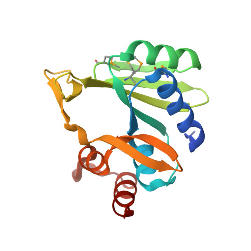 | |
UniProt | |||||
Entity Groups | |||||
| Sequence Clusters | 30% Identity50% Identity70% Identity90% Identity95% Identity100% Identity | ||||
| UniProt Group | P06107 | ||||
Sequence AnnotationsExpand | |||||
Reference Sequence | |||||
| Ligands 3 Unique | |||||
|---|---|---|---|---|---|
| ID | Chains | Name / Formula / InChI Key | 2D Diagram | 3D Interactions | |
| 3QB Download:Ideal Coordinates CCD File | F [auth A], I [auth B] | LINCOMYCIN C18 H34 N2 O6 S OJMMVQQUTAEWLP-KIDUDLJLSA-N |  | ||
| CL Download:Ideal Coordinates CCD File | C [auth A] | CHLORIDE ION Cl VEXZGXHMUGYJMC-UHFFFAOYSA-M |  | ||
| MG Download:Ideal Coordinates CCD File | D [auth A], E [auth A], G [auth B], H [auth B] | MAGNESIUM ION Mg JLVVSXFLKOJNIY-UHFFFAOYSA-N |  | ||
| Modified Residues 1 Unique | |||||
|---|---|---|---|---|---|
| ID | Chains | Type | Formula | 2D Diagram | Parent |
| MSE Query on MSE | A, B | L-PEPTIDE LINKING | C5 H11 N O2 Se |  | MET |
| Length ( Å ) | Angle ( ˚ ) |
|---|---|
| a = 56.447 | α = 90 |
| b = 63.444 | β = 101.6 |
| c = 60.808 | γ = 90 |
| Software Name | Purpose |
|---|---|
| PHENIX | refinement |
| StructureStudio | data collection |
| HKL-3000 | data reduction |
| HKL-3000 | data scaling |
| PHENIX | phasing |


