4WEG | pdb_00004weg

influenza virus neuraminidase N9 in complex 2,3-difluorosialic acid


Experimental Data Snapshot

  • Method: X-RAY DIFFRACTION
  • Resolution: 2.10 Å
  • R-Value Free: 
    0.201 (Depositor), 0.207 (DCC) 
  • R-Value Work: 
    0.143 (Depositor), 0.148 (DCC) 
  • R-Value Observed: 
    0.146 (Depositor) 

wwPDB Validation 3D Report Full Report

Validation slider image for 4WEG

Ligand Structure Quality Assessment 


This is version 2.1 of the entry. See complete history

Literature

Catalytic mechanism and novel receptor binding sites of human parainfluenza virus type 3 hemagglutinin-neuraminidase (hPIV3 HN)

Streltsov, V.A.Pilling, P.Barrett, S.McKimm-Breschkin, J.L.

(2015) Antiviral Res 123: 216-223

  • DOI: https://doi.org/10.1016/j.antiviral.2015.08.014
  • Primary Citation Related Structures: 
    4WEF, 4WEG

  • PubMed Abstract: 

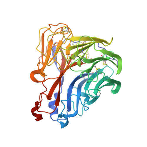
    The human parainfluenza virus type 3 (hPIV3) hemagglutinin-neuraminidase (HN) has opposing functions of binding sialic acid receptors and cleaving them, facilitating virus release. The crystal structure of hPIV3 HN complexed with the substrate analogue difluorosialic acid (DFSA) revealed that catalysis by HN involves the formation of a covalently linked sialosyl-enzyme intermediate which was trapped along with a transition-state analogue resembling an oxocarbenium ion. This mechanism of enzyme catalysis was also confirmed in the crystal structure of the influenza N9 neuraminidase complexed with DFSA. Additionally, novel secondary receptor binding sites were identified in the hPIV3 HN-DFSA complex including one near the catalytic cavity which upon binding DFSA imposes subtle changes and may help the HN balance the opposing functions. Multiple receptor binding sites may increase avidity to facilitate cell binding and fusion promotion. The secondary receptor binding sites in the paramyxoviruses are so far unique to each virus type.


  • Organizational Affiliation
    • CSIRO Manufacturing, 343 Royal Parade, Parkville, Victoria 3052, Australia. Electronic address: victor.streltsov@csiro.au.

Macromolecule Content 

  • Total Structure Weight: 46.77 kDa 
  • Atom Count: 3,720 
  • Modeled Residue Count: 388 
  • Deposited Residue Count: 388 
  • Unique protein chains: 1

Macromolecules

Find similar proteins by:|  3D Structure
Entity ID: 1
MoleculeChains  Sequence LengthOrganismDetailsImage
Neuraminidase388Influenza A virus (A/tern/Australia/G70C/1975(H11N9))Mutation(s): 0 
Gene Names: NA
EC: 3.2.1.18
UniProt
Find proteins for P03472 (Influenza A virus (strain A/Tern/Australia/G70C/1975 H11N9))
Explore P03472 
Go to UniProtKB:  P03472
Entity Groups
Sequence Clusters30% Identity50% Identity70% Identity90% Identity95% Identity100% Identity
UniProt GroupP03472
Glycosylation
Glycosylation Sites: 3
Sequence Annotations
Expand
Reference Sequence

Oligosaccharides

Help  
Entity ID: 2
MoleculeChains Length2D Diagram GlycosylationD Interactions
alpha-D-mannopyranose-(1-2)-alpha-D-mannopyranose-(1-2)-alpha-D-mannopyranose-(1-3)-[alpha-D-mannopyranose-(1-3)-alpha-D-mannopyranose-(1-6)]beta-D-mannopyranose-(1-4)-2-acetamido-2-deoxy-beta-D-glucopyranose-(1-4)-2-acetamido-2-deoxy-beta-D-glucopyranose
B
8N-Glycosylation
Glycosylation Resources
GlyTouCan: G83582BK
GlyCosmos: G83582BK
GlyGen: G83582BK
Entity ID: 3
MoleculeChains Length2D Diagram GlycosylationD Interactions
2-acetamido-2-deoxy-beta-D-glucopyranose-(1-4)-2-acetamido-2-deoxy-beta-D-glucopyranose
C
2N-Glycosylation
Glycosylation Resources
GlyTouCan: G42666HT
GlyCosmos: G42666HT
GlyGen: G42666HT

Small Molecules

Ligands 5 Unique
IDChains Name / Formula / InChI Key2D Diagram3D Interactions
SFJ

Query on SFJ


Download:Ideal Coordinates CCD File
D [auth A](2R,3R,4R,5R,6R)-5-acetamido-2,3-difluoro-4-hydroxy-6-[(1R,2R)-1,2,3-trihydroxypropyl]tetrahydro-2H-pyran-2-carboxylic acid
C11 H17 F2 N O8
HMALKZAXODOYOP-DAXAGCIGSA-N
FSI

Query on FSI


Download:Ideal Coordinates CCD File
H [auth A]5-acetamido-3,5-dideoxy-3-fluoro-D-erythro-alpha-L-manno-non-2-ulopyranosonic acid
C11 H18 F N O9
ALJLGESFXXDPKH-RISWTRDCSA-N
DF4

Query on DF4


Download:Ideal Coordinates CCD File
G [auth A](3R,4R,5R,6R)-5-(acetylamino)-3-fluoro-4-hydroxy-6-[(1R,2R)-1,2,3-trihydroxypropyl]-3,4,5,6-tetrahydropyranium-2-carboxylate
C11 H16 F N O8
OXSMZBYWMIZHJQ-LNSOWFDVSA-N
NAG

Query on NAG


Download:Ideal Coordinates CCD File
E [auth A]2-acetamido-2-deoxy-beta-D-glucopyranose
C8 H15 N O6
OVRNDRQMDRJTHS-FMDGEEDCSA-N
CA

Query on CA


Download:Ideal Coordinates CCD File
F [auth A]CALCIUM ION
Ca
BHPQYMZQTOCNFJ-UHFFFAOYSA-N

Experimental Data & Validation

Experimental Data

  • Method: X-RAY DIFFRACTION
  • Resolution: 2.10 Å
  • R-Value Free:  0.201 (Depositor), 0.207 (DCC) 
  • R-Value Work:  0.143 (Depositor), 0.148 (DCC) 
  • R-Value Observed: 0.146 (Depositor) 
Space Group: I 4 3 2
Unit Cell:
Length ( Å )Angle ( ˚ )
a = 181.194α = 90
b = 181.194β = 90
c = 181.194γ = 90
Software Package:
Software NamePurpose
REFMACrefinement
Blu-Icedata collection
HKL-2000data processing
PHASERphasing
PDB_EXTRACTdata extraction
HKLdata scaling

Structure Validation

View Full Validation Report


Ligand Structure Quality Assessment 


Entry History 

Deposition Data

Revision History  (Full details and data files)

  • Version 1.0: 2015-09-16
    Type: Initial release
  • Version 1.1: 2015-10-07
    Changes: Database references
  • Version 1.2: 2015-11-18
    Changes: Database references
  • Version 1.3: 2017-10-04
    Changes: Advisory, Data collection, Database references, Derived calculations
  • Version 2.0: 2020-07-29
    Type: Remediation
    Reason: Carbohydrate remediation
    Changes: Advisory, Atomic model, Data collection, Derived calculations, Structure summary
  • Version 2.1: 2024-11-13
    Changes: Advisory, Data collection, Database references, Derived calculations, Structure summary