4WB2 | pdb_00004wb2

Crystal structure of the mirror-image L-RNA/L-DNA aptamer NOX-D20 in complex with mouse C5a complement anaphylatoxin


Experimental Data Snapshot

  • Method: X-RAY DIFFRACTION
  • Resolution: 1.80 Å
  • R-Value Free: 
    0.193 (Depositor), 0.197 (DCC) 
  • R-Value Work: 
    0.167 (Depositor), 0.172 (DCC) 
  • R-Value Observed: 
    0.168 (Depositor) 

wwPDB Validation 3D Report Full Report

Validation slider image for 4WB2

This is version 1.2 of the entry. See complete history

Literature

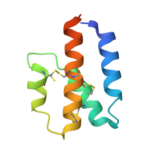
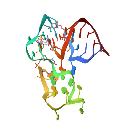
Structural basis for the targeting of complement anaphylatoxin C5a using a mixed L-RNA/L-DNA aptamer.

Yatime, L.Maasch, C.Hoehlig, K.Klussmann, S.Andersen, G.R.Vater, A.

(2015) Nat Commun 6: 6481-6481

  • DOI: https://doi.org/10.1038/ncomms7481
  • Primary Citation Related Structures: 
    4WB2, 4WB3

  • PubMed Abstract: 

    L-Oligonucleotide aptamers (Spiegelmers) consist of non-natural L-configured nucleotides and are of particular therapeutic interest due to their high resistance to plasma nucleases. The anaphylatoxin C5a, a potent inflammatory mediator generated during complement activation that has been implicated with organ damage, can be efficiently targeted by Spiegelmers. Here, we present the first crystallographic structures of an active Spiegelmer, NOX-D20, bound to its physiological targets, mouse C5a and C5a-desArg. The structures reveal a complex 3D architecture for the L-aptamer that wraps around C5a, including an intramolecular G-quadruplex stabilized by a central Ca(2+) ion. Functional validation of the observed L-aptamer:C5a binding mode through mutational studies also rationalizes the specificity of NOX-D20 for mouse and human C5a against macaque and rat C5a. Finally, our structural model provides the molecular basis for the Spiegelmer affinity improvement through positional L-ribonucleotide to L-deoxyribonucleotide exchanges and for its inhibition of the C5a:C5aR interaction.


  • Organizational Affiliation
    • Department of Molecular Biology and Genetics, Aarhus University, Gustav Wieds Vej 10C, DK-8000 Aarhus, Denmark.

Macromolecule Content 

  • Total Structure Weight: 53.7 kDa 
  • Atom Count: 4,011 
  • Modeled Residue Count: 290 
  • Deposited Residue Count: 317 
  • Unique protein chains: 1
  • Unique nucleic acid chains: 1

Macromolecules


Find similar proteins by:|  3D Structure
Entity ID: 1
MoleculeChains  Sequence LengthOrganismDetailsImage
Complement C5
A, B, C
79Mus musculusMutation(s): 0 
Gene Names: C5Hc
UniProt & NIH Common Fund Data Resources
Find proteins for P06684 (Mus musculus)
Explore P06684 
Go to UniProtKB:  P06684
IMPC:  MGI:96031
Entity Groups
Sequence Clusters30% Identity50% Identity70% Identity90% Identity95% Identity100% Identity
UniProt GroupP06684
Sequence Annotations
Expand
Reference Sequence
Find similar nucleic acids by:  (by identity cutoff) 
Entity ID: 2
MoleculeChains LengthOrganismImage
mixed L-RNA/L-DNA mirror-image aptamer NOX-D20 (40-MER)
D, E
40synthetic construct
Sequence Annotations
Expand
Reference Sequence

Small Molecules

Ligands 3 Unique
IDChains Name / Formula / InChI Key2D Diagram3D Interactions
ACT

Query on ACT


Download:Ideal Coordinates CCD File
F [auth A]
H [auth B]
I [auth B]
J [auth C]
O [auth D]
F [auth A],
H [auth B],
I [auth B],
J [auth C],
O [auth D],
S [auth E]
ACETATE ION
C2 H3 O2
QTBSBXVTEAMEQO-UHFFFAOYSA-M
CA

Query on CA


Download:Ideal Coordinates CCD File
G [auth A]
K [auth C]
L [auth D]
M [auth D]
P [auth E]
G [auth A],
K [auth C],
L [auth D],
M [auth D],
P [auth E],
Q [auth E]
CALCIUM ION
Ca
BHPQYMZQTOCNFJ-UHFFFAOYSA-N
MG

Query on MG


Download:Ideal Coordinates CCD File
N [auth D],
R [auth E]
MAGNESIUM ION
Mg
JLVVSXFLKOJNIY-UHFFFAOYSA-N

Experimental Data & Validation

Experimental Data

  • Method: X-RAY DIFFRACTION
  • Resolution: 1.80 Å
  • R-Value Free:  0.193 (Depositor), 0.197 (DCC) 
  • R-Value Work:  0.167 (Depositor), 0.172 (DCC) 
  • R-Value Observed: 0.168 (Depositor) 
Space Group: P 21 21 2
Unit Cell:
Length ( Å )Angle ( ˚ )
a = 46.47α = 90
b = 282.254β = 90
c = 45.899γ = 90
Software Package:
Software NamePurpose
PHENIXrefinement
XDSdata reduction
XSCALEdata scaling

Structure Validation

View Full Validation Report


Entry History 

Deposition Data

Revision History  (Full details and data files)

  • Version 1.0: 2015-05-06
    Type: Initial release
  • Version 1.1: 2018-01-17
    Changes: Data collection
  • Version 1.2: 2024-10-23
    Changes: Data collection, Database references, Derived calculations, Structure summary