4W8Y | pdb_00004w8y

Structure of full length Cmr2 from Pyrococcus furiosus (Manganese bound form)


Experimental Data Snapshot

  • Method: X-RAY DIFFRACTION
  • Resolution: 3.00 Å
  • R-Value Free: 
    0.259 (Depositor), 0.258 (DCC) 
  • R-Value Work: 
    0.223 (Depositor), 0.228 (DCC) 
  • R-Value Observed: 
    0.225 (Depositor) 

wwPDB Validation 3D Report Full Report

Validation slider image for 4W8Y

This is version 1.1 of the entry. See complete history

Literature

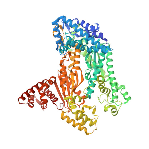
Structural Model of a CRISPR RNA-Silencing Complex Reveals the RNA-Target Cleavage Activity in Cmr4.

Benda, C.Ebert, J.Scheltema, R.A.Schiller, H.B.Baumgartner, M.Bonneau, F.Mann, M.Conti, E.

(2014) Mol Cell 56: 43-54

  • DOI: https://doi.org/10.1016/j.molcel.2014.09.002
  • Primary Citation Related Structures: 
    4W8V, 4W8W, 4W8X, 4W8Y, 4W8Z

  • PubMed Abstract: 

    The Cmr complex is an RNA-guided endonuclease that cleaves foreign RNA targets as part of the CRISPR prokaryotic defense system. We investigated the molecular architecture of the P. furiosus Cmr complex using an integrative structural biology approach. We determined crystal structures of P. furiosus Cmr1, Cmr2, Cmr4, and Cmr6 and combined them with known structural information to interpret the cryo-EM map of the complex. To support structure determination, we obtained residue-specific interaction data using protein crosslinking and mass spectrometry. The resulting pseudoatomic model reveals how the superhelical backbone of the complex is defined by the polymerizing principles of Cmr4 and Cmr5 and how it is capped at the extremities by proteins of similar folds. The inner surface of the superhelix exposes conserved residues of Cmr4 that we show are required for target-cleavage activity. The structural and biochemical data thus identify Cmr4 as the conserved endoribonuclease of the Cmr complex.


  • Organizational Affiliation
    • Department of Structural Cell Biology, Max Planck Institute of Biochemistry, 82152 Martinsried, Germany.

Macromolecule Content 

  • Total Structure Weight: 206.94 kDa 
  • Atom Count: 13,055 
  • Modeled Residue Count: 1,663 
  • Deposited Residue Count: 1,776 
  • Unique protein chains: 1

Macromolecules

Find similar proteins by:|  3D Structure
Entity ID: 1
MoleculeChains  Sequence LengthOrganismDetailsImage
CRISPR system Cmr subunit Cmr2
A, B
888Pyrococcus furiosus DSM 3638Mutation(s): 0 
Gene Names: cmr2PF1129
UniProt
Find proteins for Q8U1S6 (Pyrococcus furiosus (strain ATCC 43587 / DSM 3638 / JCM 8422 / Vc1))
Explore Q8U1S6 
Go to UniProtKB:  Q8U1S6
Entity Groups
Sequence Clusters30% Identity50% Identity70% Identity90% Identity95% Identity100% Identity
UniProt GroupQ8U1S6
Sequence Annotations
Expand
Reference Sequence

Small Molecules

Ligands 2 Unique
IDChains Name / Formula / InChI Key2D Diagram3D Interactions
ZN

Query on ZN


Download:Ideal Coordinates CCD File
C [auth A],
H [auth B]
ZINC ION
Zn
PTFCDOFLOPIGGS-UHFFFAOYSA-N
MN

Query on MN


Download:Ideal Coordinates CCD File
D [auth A]
E [auth A]
F [auth A]
G [auth A]
I [auth B]
D [auth A],
E [auth A],
F [auth A],
G [auth A],
I [auth B],
J [auth B],
K [auth B],
L [auth B]
MANGANESE (II) ION
Mn
WAEMQWOKJMHJLA-UHFFFAOYSA-N

Experimental Data & Validation

Experimental Data

  • Method: X-RAY DIFFRACTION
  • Resolution: 3.00 Å
  • R-Value Free:  0.259 (Depositor), 0.258 (DCC) 
  • R-Value Work:  0.223 (Depositor), 0.228 (DCC) 
  • R-Value Observed: 0.225 (Depositor) 
Space Group: P 1 21 1
Unit Cell:
Length ( Å )Angle ( ˚ )
a = 62.4α = 90
b = 167.925β = 98.91
c = 100.908γ = 90
Software Package:
Software NamePurpose
PHENIXrefinement
XDSdata reduction
XSCALEdata scaling

Structure Validation

View Full Validation Report


Entry History 

Deposition Data

Revision History  (Full details and data files)

  • Version 1.0: 2014-10-15
    Type: Initial release
  • Version 1.1: 2024-05-08
    Changes: Data collection, Database references, Derived calculations