4V8V | pdb_00004v8v

Structure and conformational variability of the Mycobacterium tuberculosis fatty acid synthase multienzyme complex


Experimental Data Snapshot

  • Method: ELECTRON MICROSCOPY
  • Resolution: 20.0 Å
  • Aggregation State: PARTICLE 
  • Reconstruction Method: SINGLE PARTICLE 

Starting Model: experimental
View more details

wwPDB Validation 3D Report Full Report

Validation slider image for 4V8V

This is version 2.3 of the entry. See complete history

Literature

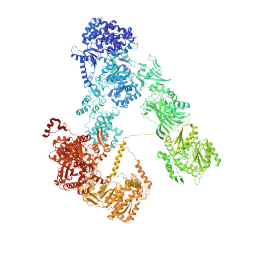
Structure and Conformational Variability of the Mycobacterium Tuberculosis Fatty Acid Synthase Multienzyme Complex.

Ciccarelli, L.Connell, S.R.Enderle, M.Mills, D.J.Vonck, J.Grininger, M.

(2013) Structure 21: 1251

  • DOI: https://doi.org/10.1016/j.str.2013.04.023
  • Primary Citation Related Structures: 
    4V8V, 4V8W

  • PubMed Abstract: 

    Antibiotic therapy in response to Mycobacterium tuberculosis infections targets de novo fatty acid biosynthesis, which is orchestrated by a 1.9 MDa type I fatty acid synthase (FAS). Here, we characterize M. tuberculosis FAS by single-particle cryo-electron microscopy and interpret the data by docking the molecular models of yeast and Mycobacterium smegmatis FAS. Our analysis reveals a porous barrel-like structure of considerable conformational variability that is illustrated by the identification of several conformational states with altered topology in the multienzymatic assembly. This demonstrates that the barrel-like structure of M. tuberculosis FAS is not just a static scaffold for the catalytic domains, but may play an active role in coordinating fatty acid synthesis. The conception of M. tuberculosis FAS as a highly dynamic assembly of domains revises the view on bacterial type I fatty acid synthesis and might inspire new strategies for inhibition of de novo fatty acid synthesis in M. tuberculosis.


  • Organizational Affiliation
    • Department of Structural Biology, Max-Planck-Institute of Biophysics, Max-von-Laue-Str. 3, 60438 Frankfurt, Germany.

Macromolecule Content 

  • Total Structure Weight: 1,982.07 kDa 
  • Atom Count: 125,856 
  • Modeled Residue Count: 16,932 
  • Deposited Residue Count: 18,534 
  • Unique protein chains: 1

Macromolecules

Find similar proteins by:|  3D Structure
Entity ID: 1
MoleculeChains  Sequence LengthOrganismDetailsImage
TYPE-I FATTY ACID SYNTHASE
A, B, C, D, E
A, B, C, D, E, F
3,089Mycobacterium tuberculosisMutation(s): 0 

Experimental Data & Validation

Experimental Data

  • Method: ELECTRON MICROSCOPY
  • Resolution: 20.0 Å
  • Aggregation State: PARTICLE 
  • Reconstruction Method: SINGLE PARTICLE 
EM Software:
TaskSoftware PackageVersion
RECONSTRUCTIONEMAN2
RECONSTRUCTIONSPARX

Structure Validation

View Full Validation Report


Entry History 

Deposition Data

Revision History  (Full details and data files)

  • Version 1.0: 2014-07-09
    Type: Initial release
  • Version 1.1: 2014-12-10
    Changes: Other
  • Version 2.0: 2017-08-02
    Changes: Advisory, Atomic model, Data collection
  • Version 2.1: 2019-12-11
    Changes: Other
  • Version 2.2: 2020-07-29
    Changes: Source and taxonomy
  • Version 2.3: 2024-05-08
    Changes: Data collection, Database references, Refinement description