4V7Q | pdb_00004v7q

Atomic model of an infectious rotavirus particle


Experimental Data Snapshot

  • Method: ELECTRON MICROSCOPY
  • Resolution: 3.80 Å
  • Aggregation State: PARTICLE 
  • Reconstruction Method: SINGLE PARTICLE 

wwPDB Validation 3D Report Full Report

Validation slider image for 4V7Q

This is version 2.1 of the entry. See complete history

Literature

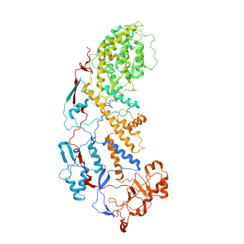
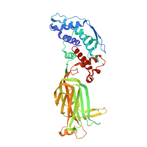
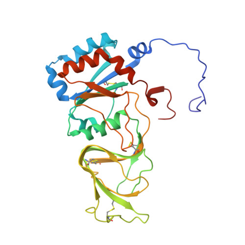
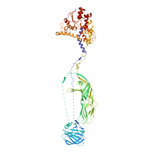
Atomic model of an infectious rotavirus particle.

Settembre, E.C.Chen, J.Z.Dormitzer, P.R.Grigorieff, N.Harrison, S.C.

(2011) EMBO J 30: 408-416

  • DOI: https://doi.org/10.1038/emboj.2010.322
  • Primary Citation Related Structures: 
    4V7Q

  • PubMed Abstract: 

    Non-enveloped viruses of different types have evolved distinct mechanisms for penetrating a cellular membrane during infection. Rotavirus penetration appears to occur by a process resembling enveloped-virus fusion: membrane distortion linked to conformational changes in a viral protein. Evidence for such a mechanism comes from crystallographic analyses of fragments of VP4, the rotavirus-penetration protein, and infectivity analyses of structure-based VP4 mutants. We describe here the structure of an infectious rotavirus particle determined by electron cryomicroscopy (cryoEM) and single-particle analysis at about 4.3 Å resolution. The cryoEM image reconstruction permits a nearly complete trace of the VP4 polypeptide chain, including the positions of most side chains. It shows how the two subfragments of VP4 (VP8(*) and VP5(*)) retain their association after proteolytic cleavage, reveals multiple structural roles for the β-barrel domain of VP5(*), and specifies interactions of VP4 with other capsid proteins. The virion model allows us to integrate structural and functional information into a coherent mechanism for rotavirus entry.


  • Organizational Affiliation
    • Laboratory of Molecular Medicine, Children's Hospital, Boston, MA, USA.

Macromolecule Content 

  • Total Structure Weight: 1,440.04 kDa 
  • Atom Count: 97,287 
  • Modeled Residue Count: 12,198 
  • Deposited Residue Count: 12,677 
  • Unique protein chains: 4

Macromolecules

Find similar proteins by:|  3D Structure
Entity ID: 1
MoleculeChains  Sequence LengthOrganismDetailsImage
Core scaffold proteinA [auth AA],
B [auth AB]
800Simian rotavirus A strain RRVMutation(s): 0 
Gene Names: Rotavirus
UniProt
Find proteins for B3F2X3 (Rotavirus A (strain RVA/Monkey/United States/RRV/1975/G3P5B[3]))
Explore B3F2X3 
Go to UniProtKB:  B3F2X3
Entity Groups
Sequence Clusters30% Identity50% Identity70% Identity90% Identity95% Identity100% Identity
UniProt GroupB3F2X3
Sequence Annotations
Expand
Reference Sequence
Find similar proteins by:|  3D Structure
Entity ID: 2
MoleculeChains  Sequence LengthOrganismDetailsImage
Intermediate capsid protein VP6397Simian rotavirus A strain RRVMutation(s): 0 
Gene Names: Rotavirus
UniProt
Find proteins for B2BN53 (Rotavirus A (strain RVA/Monkey/United States/RRV/1975/G3P5B[3]))
Explore B2BN53 
Go to UniProtKB:  B2BN53
Entity Groups
Sequence Clusters30% Identity50% Identity70% Identity90% Identity95% Identity100% Identity
UniProt GroupB2BN53
Sequence Annotations
Expand
Reference Sequence
Find similar proteins by:|  3D Structure
Entity ID: 3
MoleculeChains  Sequence LengthOrganismDetailsImage
Outer layer protein VP7276Simian rotavirus A strain RRVMutation(s): 0 
UniProt
Find proteins for P12476 (Rotavirus A (strain RVA/Monkey/United States/RRV/1975/G3P5B[3]))
Explore P12476 
Go to UniProtKB:  P12476
Entity Groups
Sequence Clusters30% Identity50% Identity70% Identity90% Identity95% Identity100% Identity
UniProt GroupP12476
Glycosylation
Glycosylation Sites: 1
Sequence Annotations
Expand
Reference Sequence
Find similar proteins by:|  3D Structure
Entity ID: 4
MoleculeChains  Sequence LengthOrganismDetailsImage
Outer capsid protein VP4CA [auth BX],
DA [auth BY],
EA [auth BZ]
776Simian rotavirus A strain RRVMutation(s): 0 
UniProt
Find proteins for C3RX20 (Rotavirus A (strain RVA/Monkey/United States/RRV/1975/G3P5B[3]))
Explore C3RX20 
Go to UniProtKB:  C3RX20
Entity Groups
Sequence Clusters30% Identity50% Identity70% Identity90% Identity95% Identity100% Identity
UniProt GroupC3RX20
Sequence Annotations
Expand
Reference Sequence

Oligosaccharides

Help  
Entity ID: 5
MoleculeChains Length2D Diagram GlycosylationD Interactions
2-acetamido-2-deoxy-beta-D-glucopyranose-(1-4)-2-acetamido-2-deoxy-beta-D-glucopyranose
FA [auth A],
GA [auth B],
HA [auth C],
IA [auth D],
JA [auth E],
FA [auth A],
GA [auth B],
HA [auth C],
IA [auth D],
JA [auth E],
KA [auth F],
LA [auth G]
2N-Glycosylation
Glycosylation Resources
GlyTouCan: G42666HT
GlyCosmos: G42666HT
GlyGen: G42666HT

Experimental Data & Validation

Experimental Data

  • Method: ELECTRON MICROSCOPY
  • Resolution: 3.80 Å
  • Aggregation State: PARTICLE 
  • Reconstruction Method: SINGLE PARTICLE 

Structure Validation

View Full Validation Report


Entry History 

Deposition Data

Revision History  (Full details and data files)

  • Version 1.0: 2014-07-09
    Type: Initial release
  • Version 1.1: 2014-12-10
    Changes: Other
  • Version 1.2: 2018-01-24
    Changes: Data collection, Structure summary
  • Version 1.3: 2019-12-18
    Changes: Database references, Derived calculations, Other
  • Version 2.0: 2020-07-29
    Type: Remediation
    Reason: Carbohydrate remediation
    Changes: Advisory, Atomic model, Data collection, Derived calculations, Structure summary
  • Version 2.1: 2024-10-16
    Changes: Data collection, Database references, Derived calculations, Structure summary