4UZZ | pdb_00004uzz

Crystal structure of the TtIFT52-46 complex


Experimental Data Snapshot

  • Method: X-RAY DIFFRACTION
  • Resolution: 2.32 Å
  • R-Value Free: 
    0.264 (Depositor), 0.260 (DCC) 
  • R-Value Work: 
    0.239 (Depositor) 
  • R-Value Observed: 
    0.241 (Depositor) 

wwPDB Validation 3D Report Full Report

Validation slider image for 4UZZ

This is version 1.1 of the entry. See complete history

Literature

Crystal Structures of Ift70/52 and Ift52/46 Provide Insight Into Intraflagellar Transport B Core Complex Assembly.

Taschner, M.Kotsis, F.Braeuer, P.Kuehn, E.W.Lorentzen, E.

(2014) J Cell Biol 207: 269

  • DOI: https://doi.org/10.1083/jcb.201408002
  • Primary Citation Related Structures: 
    4UZY, 4UZZ

  • PubMed Abstract: 

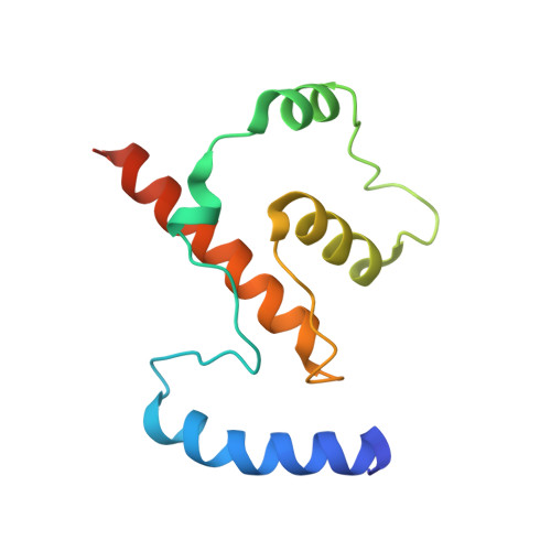
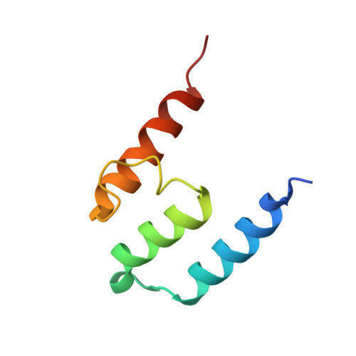
    Cilia are microtubule-based organelles that assemble via intraflagellar transport (IFT) and function as signaling hubs on eukaryotic cells. IFT relies on molecular motors and IFT complexes that mediate the contacts with ciliary cargo. To elucidate the architecture of the IFT-B complex, we reconstituted and purified the nonameric IFT-B core from Chlamydomonas reinhardtii and determined the crystal structures of C. reinhardtii IFT70/52 and Tetrahymena IFT52/46 subcomplexes. The 2.5-Å resolution IFT70/52 structure shows that IFT52330-370 is buried deeply within the IFT70 tetratricopeptide repeat superhelix. Furthermore, the polycystic kidney disease protein IFT88 binds IFT52281-329 in a complex that interacts directly with IFT70/IFT52330-381 in trans. The structure of IFT52C/IFT46C was solved at 2.3 Å resolution, and we show that it is essential for IFT-B core integrity by mediating interaction between IFT88/70/52/46 and IFT81/74/27/25/22 subcomplexes. Consistent with this, overexpression of mammalian IFT52C in MDCK cells is dominant-negative and causes IFT protein mislocalization and disrupted ciliogenesis. These data further rationalize several ciliogenesis phenotypes of IFT mutant strains.


  • Organizational Affiliation
    • Department of Structural Cell Biology, Max-Planck-Institute of Biochemistry, D-82152 Martinsried, Germany.

Macromolecule Content 

  • Total Structure Weight: 21.17 kDa 
  • Atom Count: 1,372 
  • Modeled Residue Count: 173 
  • Deposited Residue Count: 184 
  • Unique protein chains: 2

Macromolecules

Find similar proteins by:|  3D Structure
Entity ID: 1
MoleculeChains  Sequence LengthOrganismDetailsImage
INTRAFLAGELLAR TRANSPORT COMPLEX B PROTEIN 46 CARBOXY-TERMINAL PROTEIN116Tetrahymena thermophilaMutation(s): 0 
UniProt
Find proteins for Q23KH7 (Tetrahymena thermophila (strain SB210))
Explore Q23KH7 
Go to UniProtKB:  Q23KH7
Entity Groups
Sequence Clusters30% Identity50% Identity70% Identity90% Identity95% Identity100% Identity
UniProt GroupQ23KH7
Sequence Annotations
Expand
Reference Sequence
Find similar proteins by:|  3D Structure
Entity ID: 2
MoleculeChains  Sequence LengthOrganismDetailsImage
INTRAFLAGELLAR TRANSPORTER-LIKE PROTEIN68Tetrahymena thermophilaMutation(s): 0 
UniProt
Find proteins for I7LT74 (Tetrahymena thermophila (strain SB210))
Explore I7LT74 
Go to UniProtKB:  I7LT74
Entity Groups
Sequence Clusters30% Identity50% Identity70% Identity90% Identity95% Identity100% Identity
UniProt GroupI7LT74
Sequence Annotations
Expand
Reference Sequence

Experimental Data & Validation

Experimental Data

  • Method: X-RAY DIFFRACTION
  • Resolution: 2.32 Å
  • R-Value Free:  0.264 (Depositor), 0.260 (DCC) 
  • R-Value Work:  0.239 (Depositor) 
  • R-Value Observed: 0.241 (Depositor) 
Space Group: P 32 2 1
Unit Cell:
Length ( Å )Angle ( ˚ )
a = 84.394α = 90
b = 84.394β = 90
c = 95.477γ = 120
Software Package:
Software NamePurpose
PHENIXrefinement
XDSdata reduction
Aimlessdata scaling
PHENIXphasing

Structure Validation

View Full Validation Report


Entry History 

Deposition Data

Revision History  (Full details and data files)

  • Version 1.0: 2014-11-05
    Type: Initial release
  • Version 1.1: 2024-05-08
    Changes: Data collection, Database references, Other