4UZS | pdb_00004uzs

Crystal structure of Bifidobacterium bifidum beta-galactosidase


Experimental Data Snapshot

  • Method: X-RAY DIFFRACTION
  • Resolution: 1.74 Å
  • R-Value Free: 
    0.193 (Depositor), 0.191 (DCC) 
  • R-Value Work: 
    0.153 (Depositor), 0.153 (DCC) 
  • R-Value Observed: 
    0.155 (Depositor) 

wwPDB Validation 3D Report Full Report

Validation slider image for 4UZS

This is version 1.5 of the entry. See complete history

Literature

Crystal Structure of Beta1-6-Galactosidase from Bifidobacterium Bifidum S17: Trimeric Architecture, Molecular Determinants of the Enzymatic Activity and its Inhibition by Alpah-Galactose.

Godoy, A.S.Camilo, C.M.Kadowaki, M.A.Muniz, H.D.S.Santo, M.E.Murakami, M.T.Nascimento, A.S.Polikarpov, I.

(2016) FEBS J 283: 4097

  • DOI: https://doi.org/10.1111/febs.13908
  • Primary Citation Related Structures: 
    4UCF, 4UZS

  • PubMed Abstract: 

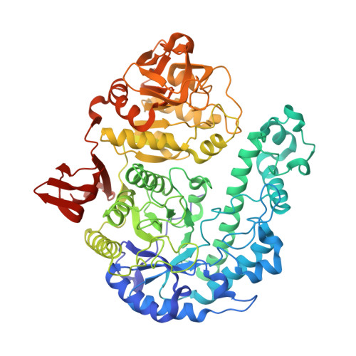
    In a search for better comprehension of β-galactosidase function and specificity, we solved the crystal structures of the GH42 β-galactosidase BbgII from Bifidobacterium bifidum S17, a well-adapted probiotic microorganism from the human digestive tract, and its complex with d-α-galactose. BbgII is a three-domain molecule that forms barrel-shaped trimers in solution. BbgII interactions with d-α-galactose, a competitive inhibitor, showed a number of residues that are involved in the coordination of ligands. A combination of site-directed mutagenesis of these amino acid residues with enzymatic activity measurements confirmed that Glu161 and Glu320 are fundamental for catalysis and their substitution by alanines led to catalytically inactive mutants. Mutation Asn160Ala resulted in a two orders of magnitude decrease of the enzyme k cat without significant modification in its K m , whereas mutations Tyr289Phe and His371Phe simultaneously decreased k cat and increased K m values. Enzymatic activity of Glu368Ala mutant was too low to be detected. Our docking and molecular dynamics simulations showed that the enzyme recognizes and tightly binds substrates with β1→6 and β1→3 bonds, while binding of the substrates with β1→4 linkages is less favorable. Structural data are available in the PDB under the accession numbers 4UZS and 4UCF.


  • Organizational Affiliation
    • Departamento de Física em São Carlos, Universidade de São Paulo, Brazil.

Macromolecule Content 

  • Total Structure Weight: 233.46 kDa 
  • Atom Count: 19,046 
  • Modeled Residue Count: 2,049 
  • Deposited Residue Count: 2,067 
  • Unique protein chains: 1

Macromolecules

Find similar proteins by:|  3D Structure
Entity ID: 1
MoleculeChains  Sequence LengthOrganismDetailsImage
BETA-GALACTOSIDASE
A, B, C
689Bifidobacterium bifidum S17Mutation(s): 0 
EC: 3.2.1.23

Small Molecules

Ligands 4 Unique
IDChains Name / Formula / InChI Key2D Diagram3D Interactions
P6G

Query on P6G


Download:Ideal Coordinates CCD File
H [auth A]HEXAETHYLENE GLYCOL
C12 H26 O7
IIRDTKBZINWQAW-UHFFFAOYSA-N
PEG

Query on PEG


Download:Ideal Coordinates CCD File
D [auth A],
G [auth A]
DI(HYDROXYETHYL)ETHER
C4 H10 O3
MTHSVFCYNBDYFN-UHFFFAOYSA-N
GOL

Query on GOL


Download:Ideal Coordinates CCD File
E [auth A]
F [auth A]
L [auth A]
M [auth B]
N [auth B]
E [auth A],
F [auth A],
L [auth A],
M [auth B],
N [auth B],
P [auth B],
Q [auth B],
R [auth C],
S [auth C]
GLYCEROL
C3 H8 O3
PEDCQBHIVMGVHV-UHFFFAOYSA-N
POL

Query on POL


Download:Ideal Coordinates CCD File
I [auth A],
J [auth A],
K [auth A],
O [auth B],
T [auth C]
N-PROPANOL
C3 H8 O
BDERNNFJNOPAEC-UHFFFAOYSA-N

Experimental Data & Validation

Experimental Data

  • Method: X-RAY DIFFRACTION
  • Resolution: 1.74 Å
  • R-Value Free:  0.193 (Depositor), 0.191 (DCC) 
  • R-Value Work:  0.153 (Depositor), 0.153 (DCC) 
  • R-Value Observed: 0.155 (Depositor) 
Space Group: P 1 21 1
Unit Cell:
Length ( Å )Angle ( ˚ )
a = 95.701α = 90
b = 99.241β = 106.47
c = 110.757γ = 90
Software Package:
Software NamePurpose
PHENIXrefinement
XDSdata reduction
SCALAdata scaling
autoSHARPphasing

Structure Validation

View Full Validation Report


Entry History 

Deposition Data

Revision History  (Full details and data files)

  • Version 1.0: 2015-09-30
    Type: Initial release
  • Version 1.1: 2016-10-12
    Changes: Database references
  • Version 1.2: 2016-12-07
    Changes: Database references
  • Version 1.3: 2017-07-05
    Changes: Data collection
  • Version 1.4: 2019-05-08
    Changes: Data collection, Experimental preparation
  • Version 1.5: 2024-05-08
    Changes: Data collection, Database references, Derived calculations, Other