4UXG | pdb_00004uxg

Crystal structure of the carboxy-terminal region of the bacteriophage T4 proximal long tail fibre protein gp34, R32 native crystal


Experimental Data Snapshot

  • Method: X-RAY DIFFRACTION
  • Resolution: 3.00 Å
  • R-Value Free: 
    0.249 (Depositor), 0.249 (DCC) 
  • R-Value Work: 
    0.226 (Depositor), 0.225 (DCC) 
  • R-Value Observed: 
    0.226 (Depositor) 

Starting Model: experimental
View more details

wwPDB Validation 3D Report Full Report

Validation slider image for 4UXG

This is version 1.3 of the entry. See complete history

Literature

Crystal Structure of the Carboxy-Terminal Region of the Bacteriophage T4 Proximal Long Tail Fiber Protein Gp34.

Granell, M.Namura, M.Alvira, S.Kanamaru, S.van Raaij, M.J.

(2017) Viruses 9

  • DOI: https://doi.org/10.3390/v9070168
  • Primary Citation Related Structures: 
    4UXE, 4UXF, 4UXG, 5NXF, 5NXH

  • PubMed Abstract: 

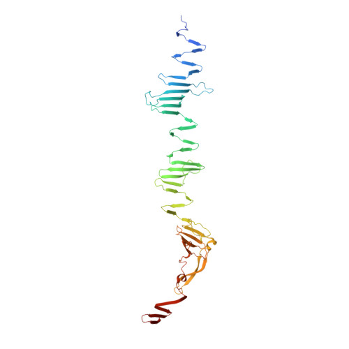
    Long tail fibers of bacteriophage T4 are formed by proteins gp34, gp35, gp36, and gp37, with gp34 located at the phage-proximal end and gp37 at the phage-distal, receptor-binding end. We have solved the structure of the carboxy-terminal region of gp34, consisting of amino acids 894-1289, by single-wavelength anomalous diffraction and extended the structure to amino acids 744-1289 using data collected from crystals containing longer gp34-fragments. The structure reveals three repeats of a mixed α-β fibrous domain in residues 744 to 877. A triple-helical neck connects to an extended triple β-helix domain (amino acids 900-1127) punctuated by two β-prism domains. Next, a β-prism domain decorated with short helices and extended β-helices is present (residues 1146-1238), while the C -terminal end is capped with another short β-helical region and three β-hairpins. The structure provides insight into the stability of the fibrous gp34 protein.


  • Organizational Affiliation
    • Departmento de Estructura de Macromoleculas, Centro Nacional de Biotecnologia (CNB-CSIC), Calle Darwin 3, E-28049 Madrid, Spain.

Macromolecule Content 

  • Total Structure Weight: 534.72 kDa 
  • Atom Count: 36,241 
  • Modeled Residue Count: 4,740 
  • Deposited Residue Count: 4,920 
  • Unique protein chains: 1

Macromolecules

Find similar proteins by:|  3D Structure
Entity ID: 1
MoleculeChains  Sequence LengthOrganismDetailsImage
LARGE TAIL FIBER PROTEIN P34
A, B, C, D, E
A, B, C, D, E, F, G, H, I, J, K, L
410Tequatrovirus T4Mutation(s): 0 
UniProt
Find proteins for P18771 (Enterobacteria phage T4)
Explore P18771 
Go to UniProtKB:  P18771
Entity Groups
Sequence Clusters30% Identity50% Identity70% Identity90% Identity95% Identity100% Identity
UniProt GroupP18771
Sequence Annotations
Expand
Reference Sequence

Experimental Data & Validation

Experimental Data

  • Method: X-RAY DIFFRACTION
  • Resolution: 3.00 Å
  • R-Value Free:  0.249 (Depositor), 0.249 (DCC) 
  • R-Value Work:  0.226 (Depositor), 0.225 (DCC) 
  • R-Value Observed: 0.226 (Depositor) 
Space Group: H 3 2
Unit Cell:
Length ( Å )Angle ( ˚ )
a = 228.489α = 90
b = 228.489β = 90
c = 1069.45γ = 120
Software Package:
Software NamePurpose
PHENIXrefinement
MOSFLMdata reduction
SCALAdata scaling
PHASERphasing

Structure Validation

View Full Validation Report


Entry History 

Deposition Data

Revision History  (Full details and data files)

  • Version 1.0: 2015-09-30
    Type: Initial release
  • Version 1.1: 2017-07-12
    Changes: Database references
  • Version 1.2: 2017-07-19
    Changes: Database references
  • Version 1.3: 2024-01-10
    Changes: Data collection, Database references, Derived calculations, Other, Refinement description