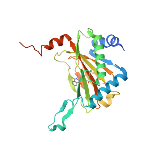
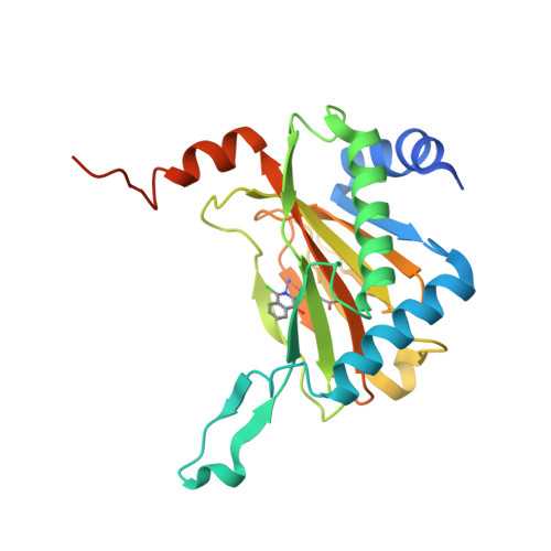
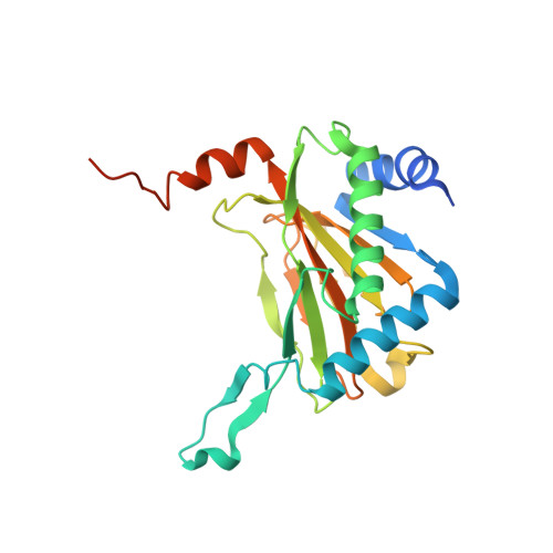
4UWD

HIF prolyl hydroxylase 2 (PHD2/ EGLN1) D315E VARIANT in complex with Mn(II) and N-[(1-chloro-4-hydroxyisoquinolin-3-yl)carbonyl]glycine (IOX3/UN9)


Experimental Data Snapshot

  • Method: X-RAY DIFFRACTION
  • Resolution: 1.72 Å
  • R-Value Free: 0.173 
  • R-Value Work: 0.149 
  • R-Value Observed: 0.151 

Starting Model: experimental
View more details

wwPDB Validation   3D Report Full Report


Ligand Structure Quality Assessment 


This is version 1.5 of the entry. See complete history


Literature

Investigating the Contribution of the Active Site Environment to the Slow Reaction of Hypoxia-Inducible Factor Prolyl Hydroxylase Domain 2 with Oxygen.

Tarhonskaya, H.Chowdhury, R.Leung, I.K.H.Loik, N.D.Mccullagh, J.S.O.Claridge, T.D.W.Schofield, C.J.Flashman, E.

(2014) Biochem J 463: 363

  • DOI: https://doi.org/10.1042/BJ20140779
  • Primary Citation of Related Structures:  
    4UWD

  • PubMed Abstract: 

    The prolyl hydroxylase domain proteins (PHDs) catalyse the post-translational hydroxylation of the hypoxia-inducible factor (HIF), a modification that regulates the hypoxic response in humans. The PHDs are Fe(II)/2-oxoglutarate (2OG) oxygenases; their catalysis is proposed to provide a link between cellular HIF levels and changes in O2 availability. Transient kinetic studies have shown that purified PHD2 reacts slowly with O2 compared with some other studied 2OG oxygenases, a property which may be related to its hypoxia-sensing role. PHD2 forms a stable complex with Fe(II) and 2OG; crystallographic and kinetic analyses indicate that an Fe(II)-co-ordinated water molecule, which must be displaced before O2 binding, is relatively stable in the active site of PHD2. We used active site substitutions to investigate whether these properties are related to the slow reaction of PHD2 with O2. While disruption of 2OG binding in a R383K variant did not accelerate O2 activation, we found that substitution of the Fe(II)-binding aspartate for a glutamate residue (D315E) manifested significantly reduced Fe(II) binding, yet maintained catalytic activity with a 5-fold faster reaction with O2. The results inform on how the precise active site environment of oxygenases can affect rates of O2 activation and provide insights into limiting steps in PHD catalysis.


  • Organizational Affiliation

    *Chemistry Research Laboratory, University of Oxford, 12 Mansfield Road, Oxford OX1 3TA, U.K.


Macromolecules
Find similar proteins by:  (by identity cutoff)  |  3D Structure
Entity ID: 1
MoleculeChains Sequence LengthOrganismDetailsImage
EGL NINE HOMOLOG 1252Homo sapiensMutation(s): 1 
EC: 1.14.11.2 (PDB Primary Data), 1.14.11 (PDB Primary Data), 1.14.11.29 (UniProt)
UniProt & NIH Common Fund Data Resources
Find proteins for Q9GZT9 (Homo sapiens)
Explore Q9GZT9 
Go to UniProtKB:  Q9GZT9
PHAROS:  Q9GZT9
GTEx:  ENSG00000135766 
Entity Groups  
Sequence Clusters30% Identity50% Identity70% Identity90% Identity95% Identity100% Identity
UniProt GroupQ9GZT9
Sequence Annotations
Expand
  • Reference Sequence
Small Molecules
Ligands 2 Unique
IDChains Name / Formula / InChI Key2D Diagram3D Interactions
UN9
Query on UN9

Download Ideal Coordinates CCD File 
C [auth A]N-[(1-CHLORO-4-HYDROXYISOQUINOLIN-3-YL)CARBONYL]GLYCINE
C12 H9 Cl N2 O4
OUQVKRKGTAUJQA-UHFFFAOYSA-N
MN
Query on MN

Download Ideal Coordinates CCD File 
B [auth A]MANGANESE (II) ION
Mn
WAEMQWOKJMHJLA-UHFFFAOYSA-N
Binding Affinity Annotations 
IDSourceBinding Affinity
UN9 BindingDB:  4UWD Kd: 80 (nM) from 1 assay(s)
IC50: min: 70, max: 7500 (nM) from 8 assay(s)
EC50: 7.90e+4 (nM) from 1 assay(s)
Experimental Data & Validation

Experimental Data

  • Method: X-RAY DIFFRACTION
  • Resolution: 1.72 Å
  • R-Value Free: 0.173 
  • R-Value Work: 0.149 
  • R-Value Observed: 0.151 
  • Space Group: P 63
Unit Cell:
Length ( Å )Angle ( ˚ )
a = 110.52α = 90
b = 110.52β = 90
c = 39.38γ = 120
Software Package:
Software NamePurpose
PHENIXrefinement
XDSdata reduction
SCALAdata scaling
PHASERphasing

Structure Validation

View Full Validation Report


Ligand Structure Quality Assessment 


Entry History 

Deposition Data

Revision History  (Full details and data files)

  • Version 1.0: 2014-08-27
    Type: Initial release
  • Version 1.1: 2014-09-10
    Changes: Atomic model, Other
  • Version 1.2: 2014-10-22
    Changes: Database references
  • Version 1.3: 2019-03-06
    Changes: Data collection, Experimental preparation, Other
  • Version 1.4: 2019-05-08
    Changes: Data collection, Experimental preparation
  • Version 1.5: 2024-01-10
    Changes: Data collection, Database references, Derived calculations, Other, Refinement description