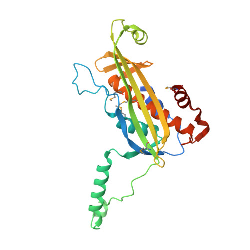
Structural and Mechanistic Insights Into the Bacterial Amyloid Secretion Channel Csgg.
Goyal, P., Krasteva, P.V., Van Gerven, N., Gubellini, F., Van Den Broeck, I., Troupiotis-Tsailaki, A., Jonckheere, W., Pehau-Arnaudet, G., Pinkner, J.S., Chapman, M.R., Hultgren, S.J., Howorka, S., Fronzes, R., Remaut, H.(2014) Nature 516: 250
- PubMed: 25219853
- DOI: https://doi.org/10.1038/nature13768
- Primary Citation of Related Structures:
4UV2, 4UV3 - PubMed Abstract:
Curli are functional amyloid fibres that constitute the major protein component of the extracellular matrix in pellicle biofilms formed by Bacteroidetes and Proteobacteria (predominantly of the α and γ classes). They provide a fitness advantage in pathogenic strains and induce a strong pro-inflammatory response during bacteraemia. Curli formation requires a dedicated protein secretion machinery comprising the outer membrane lipoprotein CsgG and two soluble accessory proteins, CsgE and CsgF. Here we report the X-ray structure of Escherichia coli CsgG in a non-lipidated, soluble form as well as in its native membrane-extracted conformation. CsgG forms an oligomeric transport complex composed of nine anticodon-binding-domain-like units that give rise to a 36-stranded β-barrel that traverses the bilayer and is connected to a cage-like vestibule in the periplasm. The transmembrane and periplasmic domains are separated by a 0.9-nm channel constriction composed of three stacked concentric phenylalanine, asparagine and tyrosine rings that may guide the extended polypeptide substrate through the secretion pore. The specificity factor CsgE forms a nonameric adaptor that binds and closes off the periplasmic face of the secretion channel, creating a 24,000 Å(3) pre-constriction chamber. Our structural, functional and electrophysiological analyses imply that CsgG is an ungated, non-selective protein secretion channel that is expected to employ a diffusion-based, entropy-driven transport mechanism.
Organizational Affiliation:
1] Structural and Molecular Microbiology, Structural Biology Research Center, VIB, Pleinlaan 2, 1050 Brussels, Belgium [2] Structural Biology Brussels, Vrije Universiteit Brussel, Pleinlaan 2, 1050 Brussels, Belgium.


