4UT1 | pdb_00004ut1

The structure of the flagellar hook junction protein FlgK from Burkholderia pseudomallei


Experimental Data Snapshot

  • Method: X-RAY DIFFRACTION
  • Resolution: 1.80 Å
  • R-Value Free: 
    0.244 (Depositor), 0.245 (DCC) 
  • R-Value Work: 
    0.198 (Depositor), 0.199 (DCC) 
  • R-Value Observed: 
    0.200 (Depositor) 

wwPDB Validation 3D Report Full Report

Validation slider image for 4UT1

This is version 1.3 of the entry. See complete history

Literature

From Crystal Structure to in Silico Epitope Discovery in Burkholderia Pseudomallei Flagellar Hook-Associated Protein Flgk.

Gourlay, L.J.Thomas, R.J.Peri, C.Conchillo-Sole, O.Ferrer-Navarro, M.Nithichanon, A.Vila, J.Daura, X.Lertmemongkolchai, G.Titball, R.Colombo, G.Bolognesi, M.

(2015) FEBS J 282: 1319

  • DOI: https://doi.org/10.1111/febs.13223
  • Primary Citation Related Structures: 
    4UT1

  • PubMed Abstract: 

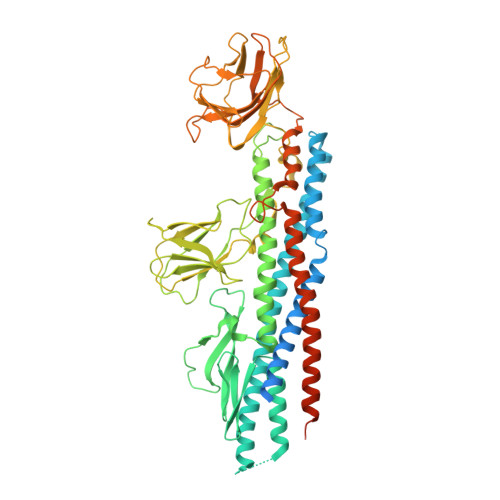
    Melioidosis, caused by the Gram-negative bacterium Burkholderia pseudomallei, is a potentially fatal infection that is endemic in Southeast Asia and Northern Australia that is poorly controlled by antibiotics. Research efforts to identify antigenic components for a melioidosis vaccine have led to the identification of several proteins, including subunits forming the flagella that mediate bacterial motility, host colonization, and virulence. This study focuses on the B. pseudomallei flagellar hook-associated protein (FlgK(Bp)), and provides the first insights into the 3D structure of FlgK proteins as targets for structure-based antigen engineering. The FlgK(Bp) crystal structure (presented here at 1.8-Å resolution) reveals a multidomain fold, comprising two small β-domains protruding from a large elongated α-helical bundle core. The evident structural similarity to flagellin, the flagellar filament subunit protein, suggests that, depending on the bacterial species, flagellar hook-associated proteins are likely to show a conserved, elongated α-helical bundle scaffold coupled to a variable number of smaller domains. Furthermore, we present immune serum recognition data confirming, in agreement with previous findings, that recovered melioidosis patients produce elevated levels of antibodies against FlgK(Bp), in comparison with seronegative and seropositive healthy subjects. Moreover, we show that FlgK(Bp) has cytotoxic effects on cultured murine macrophages, suggesting an important role in bacterial pathogenesis. Finally, computational epitope prediction methods applied to the FlgK(Bp) crystal structure, coupled with in vitro mapping, allowed us to predict three antigenic regions that locate to discrete protein domains. Taken together, our results point to FlgK(Bp) as a candidate for the design and production of epitope-containing subunits/domains as potential vaccine components.


  • Organizational Affiliation
    • Department of Biosciences, University of Milan, Italy.

Macromolecule Content 

  • Total Structure Weight: 69.74 kDa 
  • Atom Count: 4,195 
  • Modeled Residue Count: 551 
  • Deposited Residue Count: 673 
  • Unique protein chains: 1

Macromolecules

Find similar proteins by:|  3D Structure
Entity ID: 1
MoleculeChains  Sequence LengthOrganismDetailsImage
FLAGELLAR HOOK-ASSOCIATED PROTEIN673Burkholderia pseudomallei K96243Mutation(s): 0 
UniProt
Find proteins for Q63YA8 (Burkholderia pseudomallei (strain K96243))
Explore Q63YA8 
Go to UniProtKB:  Q63YA8
Entity Groups
Sequence Clusters30% Identity50% Identity70% Identity90% Identity95% Identity100% Identity
UniProt GroupQ63YA8
Sequence Annotations
Expand
Reference Sequence

Small Molecules

Ligands 1 Unique
IDChains Name / Formula / InChI Key2D Diagram3D Interactions
EDO

Query on EDO


Download:Ideal Coordinates CCD File
B [auth A]
C [auth A]
D [auth A]
E [auth A]
F [auth A]
B [auth A],
C [auth A],
D [auth A],
E [auth A],
F [auth A],
G [auth A],
H [auth A],
I [auth A],
J [auth A],
K [auth A],
L [auth A],
M [auth A],
N [auth A],
O [auth A],
P [auth A],
Q [auth A],
R [auth A],
S [auth A],
T [auth A],
U [auth A]
1,2-ETHANEDIOL
C2 H6 O2
LYCAIKOWRPUZTN-UHFFFAOYSA-N

Experimental Data & Validation

Experimental Data

  • Method: X-RAY DIFFRACTION
  • Resolution: 1.80 Å
  • R-Value Free:  0.244 (Depositor), 0.245 (DCC) 
  • R-Value Work:  0.198 (Depositor), 0.199 (DCC) 
  • R-Value Observed: 0.200 (Depositor) 
Space Group: P 1 21 1
Unit Cell:
Length ( Å )Angle ( ˚ )
a = 41.597α = 90
b = 113.005β = 106.08
c = 62.234γ = 90
Software Package:
Software NamePurpose
PHENIXrefinement
XDSdata reduction
SCALAdata scaling
BALBESphasing

Structure Validation

View Full Validation Report


Entry History 

Deposition Data

Revision History  (Full details and data files)

  • Version 1.0: 2015-02-11
    Type: Initial release
  • Version 1.1: 2015-02-18
    Changes: Database references
  • Version 1.2: 2015-04-22
    Changes: Database references
  • Version 1.3: 2024-05-08
    Changes: Data collection, Database references, Derived calculations, Other