4UST

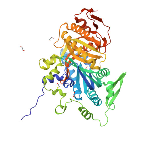
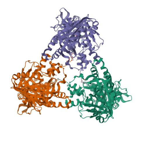
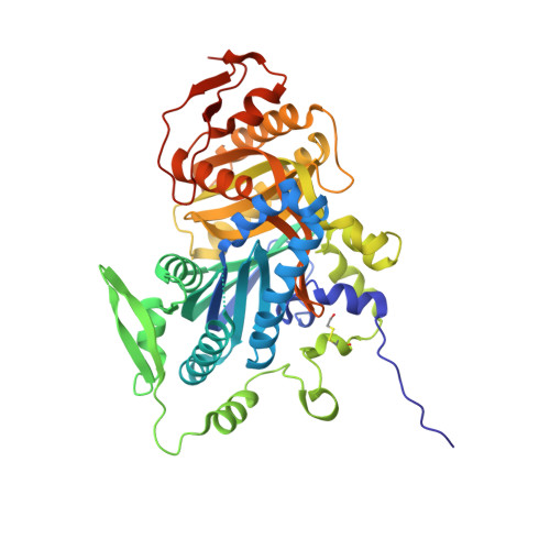
Crystal structure of human soluble Adenylyl Cyclase with pyrophosphate resulting from soaking with GTP and Magnesium


Experimental Data Snapshot

  • Method: X-RAY DIFFRACTION
  • Resolution: 1.90 Å
  • R-Value Free: 0.202 
  • R-Value Work: 0.161 
  • R-Value Observed: 0.163 

Starting Model: experimental
View more details

wwPDB Validation   3D Report Full Report


This is version 1.4 of the entry. See complete history


Literature

Structural Analysis of Human Soluble Adenylyl Cyclase and Crystal Structures of its Nucleotide Complexes -Implications for Cyclase Catalysis and Evolution.

Kleinbolting, S.Van Den Heuvel, J.Steegborn, C.

(2014) FEBS J 281: 4151

  • DOI: https://doi.org/10.1111/febs.12913
  • Primary Citation of Related Structures:  
    4UST, 4USU, 4USV, 4USW

  • PubMed Abstract: 

    The ubiquitous second messenger cAMP regulates a wide array of functions, from bacterial transcription to mammalian memory. It is synthesized by six evolutionarily distinct adenylyl cyclase (AC) families. In mammals, there are two AC types: nine transmembrane ACs (tmACs) and one soluble AC (sAC). Both AC types belong to the widespread cyclase class III, which has members in numerous organisms from archaeons to mammals. Class III also contains all known guanylyl cyclases (GCs), which synthesize the cAMP-related messenger cGMP in many eukaryotes and possibly some prokaryotes. Among mammalian ACs, sAC is uniquely regulated by bicarbonate, and has been proposed to be more closely related to a bacterial AC subfamily than to mammalian ACs, on the basis of sequence comparisons. Here, we used crystal structures of human sAC catalytic domains to analyze its relationships with other class III ACs and GCs, and to study its substrate selection mechanisms. Structural comparisons revealed a similarity within an sAC-like subfamily but no family-specific structure elements, and an unexpected sAC similarity to eukaryotic GCs and a potential bacterial GC. We further solved novel crystal structures of sAC catalytic domains in complex with a substrate analog, unprocessed ATP substrate, and product after soaking with ATP or GTP. The structures show a novel ATP-binding conformation, and suggest mechanisms for substrate association and recognition. Our results could explain the limited substrate specificity of sAC, suggest how specificity is increased in other cyclases, and indicate evolutionary relationships among class III enzymes, with sAC being close to a putative 'ancestor' cyclase. Coordinates and structure factors for the novel sAC-cat structures described have been deposited with the Worldwide PDB (www.pdb.org): ApCpp soak (entry 4usu), ATP + Ca(2+) soak (entry 4usv), GTP + Mg(2+) soak (entry 4ust), ATP soak (entry 4usw).


  • Organizational Affiliation

    Department of Biochemistry, University of Bayreuth, Germany.


Macromolecules
Find similar proteins by:  (by identity cutoff)  |  3D Structure
Entity ID: 1
MoleculeChains Sequence LengthOrganismDetailsImage
ADENYLATE CYCLASE TYPE 10475Homo sapiensMutation(s): 0 
EC: 4.6.1.1
UniProt & NIH Common Fund Data Resources
Find proteins for Q96PN6 (Homo sapiens)
Explore Q96PN6 
Go to UniProtKB:  Q96PN6
PHAROS:  Q96PN6
GTEx:  ENSG00000143199 
Entity Groups  
Sequence Clusters30% Identity50% Identity70% Identity90% Identity95% Identity100% Identity
UniProt GroupQ96PN6
Sequence Annotations
Expand
  • Reference Sequence
Small Molecules
Ligands 6 Unique
IDChains Name / Formula / InChI Key2D Diagram3D Interactions
POP
Query on POP

Download Ideal Coordinates CCD File 
E [auth A]PYROPHOSPHATE 2-
H2 O7 P2
XPPKVPWEQAFLFU-UHFFFAOYSA-L
GOL
Query on GOL

Download Ideal Coordinates CCD File 
H [auth A]GLYCEROL
C3 H8 O3
PEDCQBHIVMGVHV-UHFFFAOYSA-N
EDO
Query on EDO

Download Ideal Coordinates CCD File 
G [auth A],
I [auth A]
1,2-ETHANEDIOL
C2 H6 O2
LYCAIKOWRPUZTN-UHFFFAOYSA-N
ACT
Query on ACT

Download Ideal Coordinates CCD File 
F [auth A]ACETATE ION
C2 H3 O2
QTBSBXVTEAMEQO-UHFFFAOYSA-M
CL
Query on CL

Download Ideal Coordinates CCD File 
C [auth A],
D [auth A]
CHLORIDE ION
Cl
VEXZGXHMUGYJMC-UHFFFAOYSA-M
MG
Query on MG

Download Ideal Coordinates CCD File 
B [auth A]MAGNESIUM ION
Mg
JLVVSXFLKOJNIY-UHFFFAOYSA-N
Modified Residues  1 Unique
IDChains TypeFormula2D DiagramParent
CME
Query on CME
A
L-PEPTIDE LINKINGC5 H11 N O3 S2CYS
Experimental Data & Validation

Experimental Data

  • Method: X-RAY DIFFRACTION
  • Resolution: 1.90 Å
  • R-Value Free: 0.202 
  • R-Value Work: 0.161 
  • R-Value Observed: 0.163 
  • Space Group: P 63
Unit Cell:
Length ( Å )Angle ( ˚ )
a = 100.34α = 90
b = 100.34β = 90
c = 97.42γ = 120
Software Package:
Software NamePurpose
REFMACrefinement
XDSdata reduction
XSCALEdata scaling
MOLREPphasing

Structure Validation

View Full Validation Report


Entry History 

Deposition Data

Revision History  (Full details and data files)

  • Version 1.0: 2014-07-30
    Type: Initial release
  • Version 1.1: 2014-10-01
    Changes: Database references
  • Version 1.2: 2019-04-03
    Changes: Data collection, Derived calculations, Other, Source and taxonomy
  • Version 1.3: 2024-01-10
    Changes: Data collection, Database references, Derived calculations, Other, Refinement description
  • Version 1.4: 2024-11-20
    Changes: Structure summary