Exploring Nicotinamide Cofactor Promiscuity in Nad(P)H-Dependent Flavin Containing Monooxygenases (Fmos) Using Natural Variation within the Phosphate Binding Loop. Structure and Activity of Fmos from Cellvibrio Sp. Br and Pseudomonas Stutzeri NF13
Jensen, C.N., Ali, S.T., Allen, M.J., Grogan, G.(2014) J Mol Catal B Enzym 109: 191
- PubMed: 25383040 Search on PubMedSearch on PubMed Central
- DOI: https://doi.org/10.1016/j.molcatb.2014.08.019
- Primary Citation Related Structures:
4USQ, 4USR - PubMed Abstract:
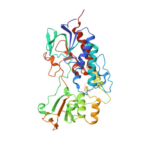
Flavin-containing monooxygenases (FMOs) catalyse asymmetric oxidation reactions that have potential for preparative organic synthesis, but most use the more expensive, phosphorylated nicotinamide cofactor NADPH to reduce FAD to FADH 2 prior to formation of the (hydro)peroxy intermediate required for substrate oxygenation. A comparison of the structures of NADPH-dependent FMO from Methylophaga aminisulfidivorans (mFMO) and SMFMO from Stenotrophomonas maltophilia , which is able to use both NADPH and NADH, suggested that the promiscuity of the latter enzyme may be due in part to the substitution of an Arg-Thr couple in the NADPH phosphate recognition site in mFMO, for a Gln-His couple in SMFMO (Jensen et al., 2012, Chembiochem , 13, 872-878). Natural variation within the phosphate binding region, and its influence on nicotinamide cofactor promiscuity, was explored through the cloning, expression, characterisation and structural studies of FMOs from Cellvibrio sp. BR (CFMO) and Pseudomonas stutzeri NF13 (PSFMO), which possess Thr-Ser and Gln-Glu in the putative phosphate recognition positions, respectively. CFMO and PSFMO displayed 5- and 1.5-fold greater activity, respectively, than SMFMO for the reduction of FAD with NADH, and were also cofactor promiscuous, displaying a ratio of activity with NADH:NADPH of 1.7:1 and 1:1.3, respectively. The structures of CFMO and PSFMO revealed the context of the phosphate binding loop in each case, and also clarified the structure of the mobile helix-loop-helix motif that appears to shield the FAD-binding pocket from bulk solvent in this class of FMOs, a feature that was absent from the structure of SMFMO.
- York Structural Biology Laboratory, Department of Chemistry, University of York, Heslington, York YO10 5DD, UK.
Organizational Affiliation: