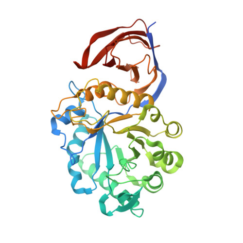
4UQC

X-ray structure of glucuronoxylan-xylanohydrolase (Xyn30A) from Clostridium thermocellum at 1.30 A resolution


Experimental Data Snapshot

  • Method: X-RAY DIFFRACTION
  • Resolution: 1.30 Å
  • R-Value Free: 0.141 
  • R-Value Work: 0.112 
  • R-Value Observed: 0.113 

Starting Model: experimental
View more details

wwPDB Validation   3D Report Full Report


Ligand Structure Quality Assessment 


This is version 1.3 of the entry. See complete history


Literature

Conservation in the Mechanism of Glucuronoxylan Hydrolysis Revealed by the Structure of Glucuronoxylan Xylano-Hydrolase (Ctxyn30A) from Clostridium Thermocellum

Freire, F.Verma, A.K.Bule, P.Alves, V.D.Fontes, C.M.G.A.Goyal, A.Najmudin, S.

(2016) Acta Crystallogr D Biol Crystallogr 72: 1162

  • DOI: https://doi.org/10.1107/S2059798316014376
  • Primary Citation of Related Structures:  
    4CKQ, 4UQ9, 4UQA, 4UQB, 4UQC, 4UQD, 4UQE, 5A6L, 5A6M

  • PubMed Abstract: 

    Glucuronoxylan endo-β-1,4-xylanases cleave the xylan chain specifically at sites containing 4-O-methylglucuronic acid substitutions. These enzymes have recently received considerable attention owing to their importance in the cooperative hydrolysis of heteropolysaccharides. However, little is known about the hydrolysis of glucuronoxylans in extreme environments. Here, the structure of a thermostable family 30 glucuronoxylan endo-β-1,4-xylanase (CtXyn30A) from Clostridium thermocellum is reported. CtXyn30A is part of the cellulosome, a highly elaborate multi-enzyme complex secreted by the bacterium to efficiently deconstruct plant cell-wall carbohydrates. CtXyn30A preferably hydrolyses glucuronoxylans and displays maximum activity at pH 6.0 and 70°C. The structure of CtXyn30A displays a (β/α) 8 TIM-barrel core with a side-associated β-sheet domain. Structural analysis of the CtXyn30A mutant E225A, solved in the presence of xylotetraose, revealed xylotetraose-cleavage oligosaccharides partially occupying subsites -3 to +2. The sugar ring at the +1 subsite is held in place by hydrophobic stacking interactions between Tyr139 and Tyr200 and hydrogen bonds to the OH group of Tyr227. Although family 30 glycoside hydrolases are retaining enzymes, the xylopyranosyl ring at the -1 subsite of CtXyn30A-E225A appears in the α-anomeric configuration. A set of residues were found to be strictly conserved in glucuronoxylan endo-β-1,4-xylanases and constitute the molecular determinants of the restricted specificity displayed by these enzymes. CtXyn30A is the first thermostable glucuronoxylan endo-β-1,4-xylanase described to date. This work reveals that substrate recognition by both thermophilic and mesophilic glucuronoxylan endo-β-1,4-xylanases is modulated by a conserved set of residues.


  • Organizational Affiliation

    CIISA-Faculdade de Medicina Veterinária, Universidade de Lisboa, Avenida da Universidade Técnica, 1300-477 Lisboa, Portugal.


Macromolecules
Find similar proteins by:  (by identity cutoff)  |  3D Structure
Entity ID: 1
MoleculeChains Sequence LengthOrganismDetailsImage
CARBOHYDRATE BINDING FAMILY 6409Acetivibrio thermocellusMutation(s): 2 
EC: 3.2.1.8
UniProt
Find proteins for A3DJS9 (Acetivibrio thermocellus (strain ATCC 27405 / DSM 1237 / JCM 9322 / NBRC 103400 / NCIMB 10682 / NRRL B-4536 / VPI 7372))
Explore A3DJS9 
Go to UniProtKB:  A3DJS9
Entity Groups  
Sequence Clusters30% Identity50% Identity70% Identity90% Identity95% Identity100% Identity
UniProt GroupA3DJS9
Sequence Annotations
Expand
  • Reference Sequence
Small Molecules
Ligands 3 Unique
IDChains Name / Formula / InChI Key2D Diagram3D Interactions
GLC
Query on GLC

Download Ideal Coordinates CCD File 
D [auth A]alpha-D-glucopyranose
C6 H12 O6
WQZGKKKJIJFFOK-DVKNGEFBSA-N
TLA
Query on TLA

Download Ideal Coordinates CCD File 
C [auth A]L(+)-TARTARIC ACID
C4 H6 O6
FEWJPZIEWOKRBE-JCYAYHJZSA-N
TAR
Query on TAR

Download Ideal Coordinates CCD File 
B [auth A]D(-)-TARTARIC ACID
C4 H6 O6
FEWJPZIEWOKRBE-LWMBPPNESA-N
Experimental Data & Validation

Experimental Data

  • Method: X-RAY DIFFRACTION
  • Resolution: 1.30 Å
  • R-Value Free: 0.141 
  • R-Value Work: 0.112 
  • R-Value Observed: 0.113 
  • Space Group: P 1
Unit Cell:
Length ( Å )Angle ( ˚ )
a = 50.39α = 111.9
b = 50.44β = 110.8
c = 58.19γ = 97.67
Software Package:
Software NamePurpose
REFMACrefinement
XDSdata reduction
XDSdata scaling
PHASERphasing

Structure Validation

View Full Validation Report


Ligand Structure Quality Assessment 


Entry History 

Deposition Data

Revision History  (Full details and data files)

  • Version 1.0: 2015-06-24
    Type: Initial release
  • Version 1.1: 2016-11-23
    Changes: Database references
  • Version 1.2: 2020-07-29
    Type: Remediation
    Reason: Carbohydrate remediation
    Changes: Data collection, Derived calculations, Other, Structure summary
  • Version 1.3: 2024-01-10
    Changes: Data collection, Database references, Refinement description, Structure summary