4UOR | pdb_00004uor

Structure of lipoteichoic acid synthase LtaS from Listeria monocytogenes in complex with glycerol phosphate


Experimental Data Snapshot

  • Method: X-RAY DIFFRACTION
  • Resolution: 2.19 Å
  • R-Value Free: 
    0.214 (Depositor), 0.215 (DCC) 
  • R-Value Work: 
    0.178 (Depositor), 0.180 (DCC) 
  • R-Value Observed: 
    0.180 (Depositor) 

Starting Model: experimental
View more details

wwPDB Validation 3D Report Full Report

Validation slider image for 4UOR

Ligand Structure Quality Assessment 


This is version 1.5 of the entry. See complete history

Literature

Structural and mechanistic insight into the Listeria monocytogenes two-enzyme lipoteichoic acid synthesis system.

Campeotto, I.Percy, M.G.MacDonald, J.T.Forster, A.Freemont, P.S.Grundling, A.

(2014) J Biological Chem 289: 28054-28069

  • DOI: https://doi.org/10.1074/jbc.M114.590570
  • Primary Citation Related Structures: 
    4UOO, 4UOP, 4UOR

  • PubMed Abstract: 

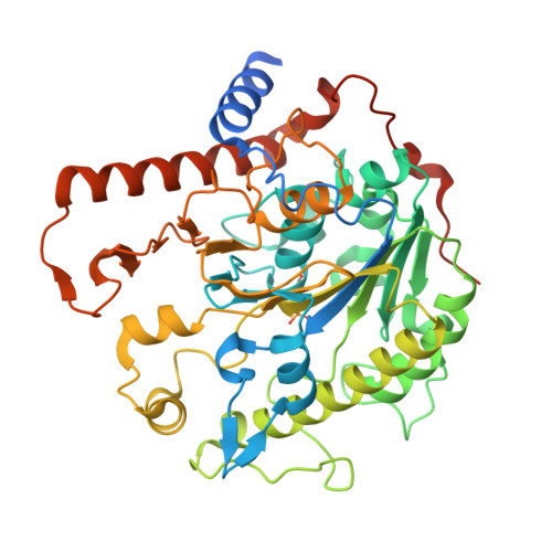
    Lipoteichoic acid (LTA) is an important cell wall component required for proper cell growth in many Gram-positive bacteria. In Listeria monocytogenes, two enzymes are required for the synthesis of this polyglycerolphosphate polymer. The LTA primase LtaP(Lm) initiates LTA synthesis by transferring the first glycerolphosphate (GroP) subunit onto the glycolipid anchor and the LTA synthase LtaS(Lm) extends the polymer by the repeated addition of GroP subunits to the tip of the growing chain. Here, we present the crystal structures of the enzymatic domains of LtaP(Lm) and LtaS(Lm). Although the enzymes share the same fold, substantial differences in the cavity of the catalytic site and surface charge distribution contribute to enzyme specialization. The eLtaS(Lm) structure was also determined in complex with GroP revealing a second GroP binding site. Mutational analysis confirmed an essential function for this binding site and allowed us to propose a model for the binding of the growing chain.


  • Organizational Affiliation
    • From the Section of Microbiology and MRC Centre for Molecular Bacteriology and Infection, and.

Macromolecule Content 

  • Total Structure Weight: 579.13 kDa 
  • Atom Count: 40,238 
  • Modeled Residue Count: 4,591 
  • Deposited Residue Count: 5,049 
  • Unique protein chains: 1

Macromolecules

Find similar proteins by:|  3D Structure
Entity ID: 1
MoleculeChains  Sequence LengthOrganismDetailsImage
LIPOTEICHOIC ACID SYNTHASE
A, B, C, D, E
A, B, C, D, E, F, G, H, I, J, K
459Listeria monocytogenes EGD-eMutation(s): 0 
EC: 2.7.8
UniProt
Find proteins for Q8Y8H6 (Listeria monocytogenes serovar 1/2a (strain ATCC BAA-679 / EGD-e))
Explore Q8Y8H6 
Go to UniProtKB:  Q8Y8H6
Entity Groups
Sequence Clusters30% Identity50% Identity70% Identity90% Identity95% Identity100% Identity
UniProt GroupQ8Y8H6
Sequence Annotations
Expand
Reference Sequence

Small Molecules

Ligands 2 Unique
IDChains Name / Formula / InChI Key2D Diagram3D Interactions
GP9

Query on GP9


Download:Ideal Coordinates CCD File
AA [auth H]
CA [auth I]
EA [auth J]
GA [auth K]
M [auth A]
AA [auth H],
CA [auth I],
EA [auth J],
GA [auth K],
M [auth A],
O [auth B],
Q [auth C],
S [auth D],
U [auth E],
W [auth F],
Y [auth G]
(2R)-2,3-dihydroxypropyl phosphate
C3 H7 O6 P
AWUCVROLDVIAJX-GSVOUGTGSA-L
MG

Query on MG


Download:Ideal Coordinates CCD File
BA [auth I]
DA [auth J]
FA [auth K]
L [auth A]
N [auth B]
BA [auth I],
DA [auth J],
FA [auth K],
L [auth A],
N [auth B],
P [auth C],
R [auth D],
T [auth E],
V [auth F],
X [auth G],
Z [auth H]
MAGNESIUM ION
Mg
JLVVSXFLKOJNIY-UHFFFAOYSA-N
Modified Residues  1 Unique
IDChains TypeFormula2D DiagramParent
TPO
Query on TPO
A, B, C, D, E
A, B, C, D, E, F, G, H, I, J, K
L-PEPTIDE LINKINGC4 H10 N O6 PTHR

Experimental Data & Validation

Experimental Data

  • Method: X-RAY DIFFRACTION
  • Resolution: 2.19 Å
  • R-Value Free:  0.214 (Depositor), 0.215 (DCC) 
  • R-Value Work:  0.178 (Depositor), 0.180 (DCC) 
  • R-Value Observed: 0.180 (Depositor) 
Space Group: P 21 21 21
Unit Cell:
Length ( Å )Angle ( ˚ )
a = 119.246α = 90
b = 119.625β = 90
c = 472.658γ = 90
Software Package:
Software NamePurpose
PHENIXrefinement
XDSdata reduction
SCALAdata scaling
PHASERphasing

Structure Validation

View Full Validation Report


Ligand Structure Quality Assessment 


Entry History 

Deposition Data

Revision History  (Full details and data files)

  • Version 1.0: 2014-08-27
    Type: Initial release
  • Version 1.1: 2014-09-03
    Changes: Structure summary
  • Version 1.2: 2014-10-22
    Changes: Database references
  • Version 1.3: 2018-02-28
    Changes: Database references
  • Version 1.4: 2024-01-10
    Changes: Data collection, Database references, Derived calculations, Other, Refinement description
  • Version 1.5: 2024-11-06
    Changes: Structure summary