4UMK | pdb_00004umk

The complex of Spo0J and parS DNA in chromosomal partition system


Experimental Data Snapshot

  • Method: X-RAY DIFFRACTION
  • Resolution: 3.10 Å
  • R-Value Free: 
    0.299 (Depositor), 0.301 (DCC) 
  • R-Value Work: 
    0.251 (Depositor), 0.252 (DCC) 
  • R-Value Observed: 
    0.254 (Depositor) 

wwPDB Validation 3D Report Full Report

Validation slider image for 4UMK

This is version 1.2 of the entry. See complete history

Literature

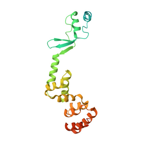
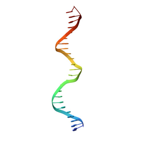
Insights into ParB spreading from the complex structure of Spo0J and parS.

Chen, B.W.Lin, M.H.Chu, C.H.Hsu, C.E.Sun, Y.J.

(2015) Proc Natl Acad Sci U S A 112: 6613-6618

  • DOI: https://doi.org/10.1073/pnas.1421927112
  • Primary Citation Related Structures: 
    4UMK

  • PubMed Abstract: 

    Spo0J (stage 0 sporulation protein J, a member of the ParB superfamily) is an essential component of the ParABS (partition system of ParA, ParB, and parS)-related bacterial chromosome segregation system. ParB (partition protein B) and its regulatory protein, ParA, act cooperatively through parS (partition S) DNA to facilitate chromosome segregation. ParB binds to chromosomal DNA at specific parS sites as well as the neighboring nonspecific DNA sites. Various ParB molecules can associate together and spread along the chromosomal DNA. ParB oligomer and parS DNA interact together to form a high-order nucleoprotein that is required for the loading of the structural maintenance of chromosomes proteins onto the chromosome for chromosomal DNA condensation. In this report, we characterized the binding of parS and Spo0J from Helicobacter pylori (HpSpo0J) and solved the crystal structure of the C-terminal domain truncated protein (Ct-HpSpo0J)-parS complex. Ct-HpSpo0J folds into an elongated structure that includes a flexible N-terminal domain for protein-protein interaction and a conserved DNA-binding domain for parS binding. Two Ct-HpSpo0J molecules bind with one parS. Ct-HpSpo0J interacts vertically and horizontally with its neighbors through the N-terminal domain to form an oligomer. These adjacent and transverse interactions are accomplished via a highly conserved arginine patch: RRLR. These interactions might be needed for molecular assembly of a high-order nucleoprotein complex and for ParB spreading. A structural model for ParB spreading and chromosomal DNA condensation that lead to chromosome segregation is proposed.


  • Organizational Affiliation
    • Department of Life Science and Institute of Bioinformatics and Structural Biology, College of Life Science, National Tsing Hua University, Hsinchu 30013, Taiwan.

Macromolecule Content 

  • Total Structure Weight: 139.59 kDa 
  • Atom Count: 7,994 
  • Modeled Residue Count: 820 
  • Deposited Residue Count: 1,056 
  • Unique protein chains: 1
  • Unique nucleic acid chains: 2

Macromolecules


Find similar proteins by:|  3D Structure
Entity ID: 1
MoleculeChains  Sequence LengthOrganismDetailsImage
PROBABLE CHROMOSOME-PARTITIONING PROTEIN PARB
A, B, C, D
240Helicobacter pylori 26695Mutation(s): 0 
UniProt
Find proteins for O25758 (Helicobacter pylori (strain ATCC 700392 / 26695))
Explore O25758 
Go to UniProtKB:  O25758
Entity Groups
Sequence Clusters30% Identity50% Identity70% Identity90% Identity95% Identity100% Identity
UniProt GroupO25758
Sequence Annotations
Expand
Reference Sequence
Find similar nucleic acids by:  Sequence
Entity ID: 2
MoleculeChains LengthOrganismImage
DNAE [auth W],
H [auth Z]
24Bacillus subtilis
Sequence Annotations
Expand
Reference Sequence
Find similar nucleic acids by:  Sequence
Entity ID: 3
MoleculeChains LengthOrganismImage
DNAF [auth X],
G [auth Y]
24Bacillus subtilis
Sequence Annotations
Expand
Reference Sequence

Experimental Data & Validation

Experimental Data

  • Method: X-RAY DIFFRACTION
  • Resolution: 3.10 Å
  • R-Value Free:  0.299 (Depositor), 0.301 (DCC) 
  • R-Value Work:  0.251 (Depositor), 0.252 (DCC) 
  • R-Value Observed: 0.254 (Depositor) 
Space Group: P 1 21 1
Unit Cell:
Length ( Å )Angle ( ˚ )
a = 54.475α = 90
b = 232.731β = 109.15
c = 78.342γ = 90
Software Package:
Software NamePurpose
PHENIXrefinement
HKL-2000data reduction
HKL-2000data scaling

Structure Validation

View Full Validation Report


Entry History 

Deposition Data

Revision History  (Full details and data files)

  • Version 1.0: 2015-05-13
    Type: Initial release
  • Version 1.1: 2018-08-29
    Changes: Data collection, Database references
  • Version 1.2: 2024-05-08
    Changes: Data collection, Database references, Derived calculations, Other