4UJ3 | pdb_00004uj3

Crystal structure of human Rab11-Rabin8-FIP3


Experimental Data Snapshot

  • Method: X-RAY DIFFRACTION
  • Resolution: 3.00 Å
  • R-Value Free: 
    0.250 (Depositor), 0.251 (DCC) 
  • R-Value Work: 
    0.198 (Depositor), 0.202 (DCC) 
  • R-Value Observed: 
    0.201 (Depositor) 

Starting Model: experimental
View more details

wwPDB Validation 3D Report Full Report

Validation slider image for 4UJ3

Ligand Structure Quality Assessment 


This is version 1.3 of the entry. See complete history

Literature

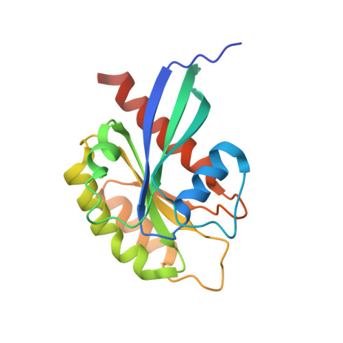
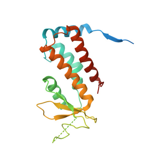
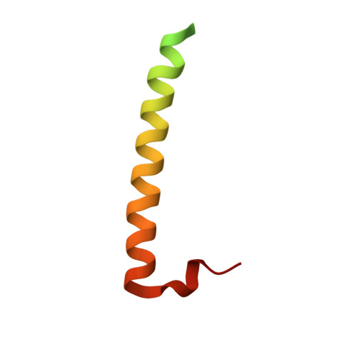
Structure of Rab11-Fip3-Rabin8 Reveals Simultaneous Binding of Fip3 and Rabin8 Effectors to Rab11.

Vetter, M.Stehle, R.Basquin, C.Lorentzen, E.

(2015) Nat Struct Mol Biol 22: 695

  • DOI: https://doi.org/10.1038/nsmb.3065
  • Primary Citation Related Structures: 
    4UJ3, 4UJ4, 4UJ5

  • PubMed Abstract: 

    The small GTPase Rab11 and its effectors FIP3 and Rabin8 are essential to membrane-trafficking pathways required for cytokinesis and ciliogenesis. Although effector binding is generally assumed to be sequential and mutually exclusive, we show that Rab11 can simultaneously bind FIP3 and Rabin8. We determined crystal structures of human Rab11-GMPPNP-Rabin8 and Rab11-GMPPNP-FIP3-Rabin8. The structures reveal that the C-terminal domain of Rabin8 adopts a previously undescribed fold that interacts with Rab11 at an unusual effector-binding site neighboring the canonical FIP3-binding site. We show that Rab11-GMPPNP-FIP3-Rabin8 is more stable than Rab11-GMPPNP-Rabin8, owing to direct interaction between Rabin8 and FIP3 within the dual effector-bound complex. The data allow us to propose a model for how membrane-targeting complexes assemble at the trans-Golgi network and recycling endosomes, through multiple weak interactions that create high-avidity complexes.


  • Organizational Affiliation
    • Department of Structural Cell Biology, Max Planck Institute of Biochemistry, Martinsried, Germany.

Macromolecule Content 

  • Total Structure Weight: 409.49 kDa 
  • Atom Count: 23,374 
  • Modeled Residue Count: 2,965 
  • Deposited Residue Count: 3,584 
  • Unique protein chains: 3

Macromolecules

Find similar proteins by:|  3D Structure
Entity ID: 1
MoleculeChains  Sequence LengthOrganismDetailsImage
RAS-RELATED PROTEIN RAB-11A
A, D, G, J, M
A, D, G, J, M, P, S, V
187Homo sapiensMutation(s): 1 
EC: 3.6.5.2
UniProt & NIH Common Fund Data Resources
Find proteins for P62491 (Homo sapiens)
Explore P62491 
Go to UniProtKB:  P62491
PHAROS:  P62491
GTEx:  ENSG00000103769 
Entity Groups
Sequence Clusters30% Identity50% Identity70% Identity90% Identity95% Identity100% Identity
UniProt GroupP62491
Sequence Annotations
Expand
Reference Sequence
Find similar proteins by:|  3D Structure
Entity ID: 2
MoleculeChains  Sequence LengthOrganismDetailsImage
RAB-3A-INTERACTING PROTEIN
B, E, H, K, N
B, E, H, K, N, Q, T, W
195Homo sapiensMutation(s): 0 
UniProt & NIH Common Fund Data Resources
Find proteins for Q96QF0 (Homo sapiens)
Explore Q96QF0 
Go to UniProtKB:  Q96QF0
PHAROS:  Q96QF0
GTEx:  ENSG00000127328 
Entity Groups
Sequence Clusters30% Identity50% Identity70% Identity90% Identity95% Identity100% Identity
UniProt GroupQ96QF0
Sequence Annotations
Expand
Reference Sequence
Find similar proteins by:|  3D Structure
Entity ID: 3
MoleculeChains  Sequence LengthOrganismDetailsImage
RAB11 FAMILY-INTERACTING PROTEIN 3
C, F, I, L, O
C, F, I, L, O, R, U, X
66Homo sapiensMutation(s): 0 
UniProt & NIH Common Fund Data Resources
Find proteins for O75154 (Homo sapiens)
Explore O75154 
Go to UniProtKB:  O75154
PHAROS:  O75154
GTEx:  ENSG00000090565 
Entity Groups
Sequence Clusters30% Identity50% Identity70% Identity90% Identity95% Identity100% Identity
UniProt GroupO75154
Sequence Annotations
Expand
Reference Sequence

Small Molecules

Ligands 4 Unique
IDChains Name / Formula / InChI Key2D Diagram3D Interactions
GNP

Query on GNP


Download:Ideal Coordinates CCD File
AA [auth D]
CA [auth G]
EA [auth J]
GA [auth M]
JA [auth P]
AA [auth D],
CA [auth G],
EA [auth J],
GA [auth M],
JA [auth P],
LA [auth S],
NA [auth V],
Y [auth A]
PHOSPHOAMINOPHOSPHONIC ACID-GUANYLATE ESTER
C10 H17 N6 O13 P3
UQABYHGXWYXDTK-UUOKFMHZSA-N
PG4

Query on PG4


Download:Ideal Coordinates CCD File
PA [auth V]TETRAETHYLENE GLYCOL
C8 H18 O5
UWHCKJMYHZGTIT-UHFFFAOYSA-N
SO4

Query on SO4


Download:Ideal Coordinates CCD File
IA [auth M]SULFATE ION
O4 S
QAOWNCQODCNURD-UHFFFAOYSA-L
MG

Query on MG


Download:Ideal Coordinates CCD File
BA [auth D]
DA [auth G]
FA [auth J]
HA [auth M]
KA [auth P]
BA [auth D],
DA [auth G],
FA [auth J],
HA [auth M],
KA [auth P],
MA [auth S],
OA [auth V],
Z [auth A]
MAGNESIUM ION
Mg
JLVVSXFLKOJNIY-UHFFFAOYSA-N

Experimental Data & Validation

Experimental Data

  • Method: X-RAY DIFFRACTION
  • Resolution: 3.00 Å
  • R-Value Free:  0.250 (Depositor), 0.251 (DCC) 
  • R-Value Work:  0.198 (Depositor), 0.202 (DCC) 
  • R-Value Observed: 0.201 (Depositor) 
Space Group: P 1 21 1
Unit Cell:
Length ( Å )Angle ( ˚ )
a = 67.498α = 90
b = 165.374β = 95.93
c = 218.687γ = 90
Software Package:
Software NamePurpose
PHENIXrefinement
XDSdata reduction
Aimlessdata scaling
PHASERphasing

Structure Validation

View Full Validation Report


Ligand Structure Quality Assessment 


Entry History 

Deposition Data

Revision History  (Full details and data files)

  • Version 1.0: 2015-08-12
    Type: Initial release
  • Version 1.1: 2015-08-19
    Changes: Database references
  • Version 1.2: 2015-09-16
    Changes: Database references
  • Version 1.3: 2024-01-10
    Changes: Data collection, Database references, Derived calculations, Other, Refinement description