4UIF | pdb_00004uif

Cryo-EM structure of Dengue virus serotype 2 in complex with antigen-binding fragments of human antibody 2D22


Experimental Data Snapshot

  • Method: ELECTRON MICROSCOPY
  • Resolution: 6.50 Å
  • Aggregation State: PARTICLE 
  • Reconstruction Method: SINGLE PARTICLE 

Starting Model: experimental
View more details

wwPDB Validation 3D Report Full Report

Validation slider image for 4UIF

This is version 1.3 of the entry. See complete history. 

Literature

Cryo-EM structure of an antibody that neutralizes dengue virus type 2 by locking E protein dimers.

Fibriansah, G., Ibarra, K.D., Ng, T.S., Smith, S.A., Tan, J.L., Lim, X.N., Ooi, J.S., Kostyuchenko, V.A., Wang, J., de Silva, A.M., Harris, E., Crowe, J.E., Lok, S.M.

(2015) Science 349: 88-91

  • DOI: https://doi.org/10.1126/science.aaa8651
  • Primary Citation Related Structures: 
    4UIF, 4UIH, 5A1Z

  • PubMed Abstract: 

    There are four closely-related dengue virus (DENV) serotypes. Infection with one serotype generates antibodies that may cross-react and enhance infection with other serotypes in a secondary infection. We demonstrated that DENV serotype 2 (DENV2)-specific human monoclonal antibody (HMAb) 2D22 is therapeutic in a mouse model of antibody-enhanced severe dengue disease. We determined the cryo-electron microscopy (cryo-EM) structures of HMAb 2D22 complexed with two different DENV2 strains. HMAb 2D22 binds across viral envelope (E) proteins in the dimeric structure, which probably blocks the E protein reorganization required for virus fusion. HMAb 2D22 "locks" two-thirds of or all dimers on the virus surface, depending on the strain, but neutralizes these DENV2 strains with equal potency. The epitope defined by HMAb 2D22 is a potential target for vaccines and therapeutics.


  • Organizational Affiliation: 
    • Program in Emerging Infectious Diseases, Duke-National University of Singapore Graduate Medical School, Singapore. Centre for BioImaging Sciences, National University of Singapore, Singapore.

Macromolecule Content 

  • Total Structure Weight: 264.79 kDa 
  • Atom Count: 2,394 
  • Modeled Residue Count: 2,394 
  • Deposited Residue Count: 2,430 
  • Unique protein chains: 4

Macromolecules

Find similar proteins by:|  3D Structure
Entity ID: 1
MoleculeChains  Sequence LengthOrganismDetailsImage
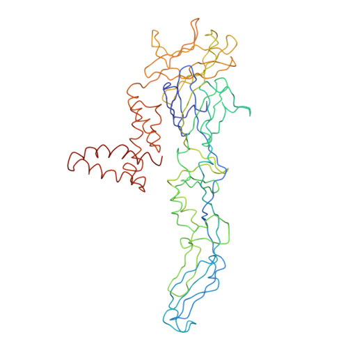
DENGUE VIRUS SEROTYPE 2 STRAIN PVP94 07 - ENVELOPE PROTEIN
A, C, E
495dengue virus type 2Mutation(s): 0 
EC: 2.7.7.48
UniProt
Find proteins for E0WXJ2 (Dengue virus type 2)
Explore E0WXJ2 
Go to UniProtKB:  E0WXJ2
Entity Groups
Sequence Clusters30% Identity50% Identity70% Identity90% Identity95% Identity100% Identity
UniProt GroupE0WXJ2
Sequence Annotations
Expand
Reference Sequence
Find similar proteins by:|  3D Structure
Entity ID: 2
MoleculeChains  Sequence LengthOrganismDetailsImage
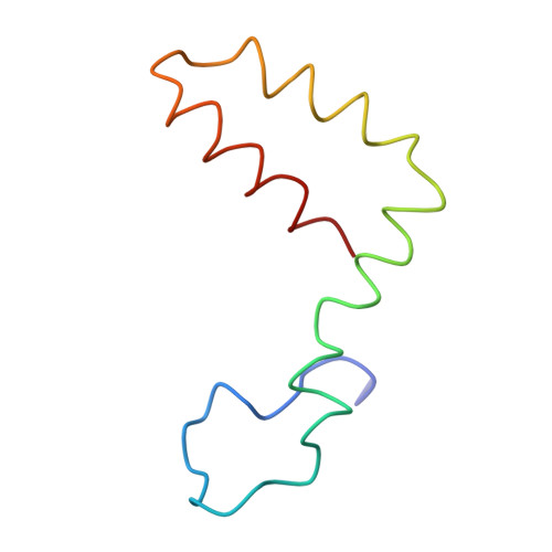
DENGUE VIRUS SEROTYPE 2 STRAIN STRAIN PVP94 07 - MEMBRANE PROTEIN
B, D, F
72dengue virus type 2Mutation(s): 0 
EC: 2.7.7.48
UniProt
Find proteins for E0WXJ2 (Dengue virus type 2)
Explore E0WXJ2 
Go to UniProtKB:  E0WXJ2
Entity Groups
Sequence Clusters30% Identity50% Identity70% Identity90% Identity95% Identity100% Identity
UniProt GroupE0WXJ2
Sequence Annotations
Expand
Reference Sequence
Find similar proteins by:|  3D Structure
Entity ID: 3
MoleculeChains  Sequence LengthOrganismDetailsImage
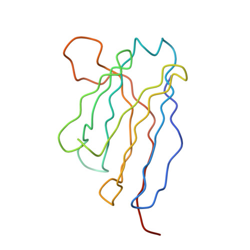
ANTIGEN-BINDING FRAGMENT OF HUMAN ANTIBODY 2D22 - HEAVY CHAIN
G, I, K
128Homo sapiensMutation(s): 0 
Entity Groups
Sequence Clusters30% Identity50% Identity70% Identity90% Identity95% Identity100% Identity
Sequence Annotations
Expand
Reference Sequence
Find similar proteins by:|  3D Structure
Entity ID: 4
MoleculeChains  Sequence LengthOrganismDetailsImage
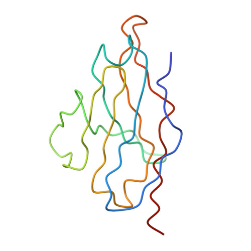
ANTIGEN-BINDING FRAGMENT OF HUMAN ANTIBODY 2D22 - LIGHT CHAIN
H, J, L
115Homo sapiensMutation(s): 0 
UniProt
Find proteins for S6BGD6 (Homo sapiens)
Explore S6BGD6 
Go to UniProtKB:  S6BGD6
Entity Groups
Sequence Clusters30% Identity50% Identity70% Identity90% Identity95% Identity100% Identity
UniProt GroupS6BGD6
Sequence Annotations
Expand
Reference Sequence

Experimental Data & Validation

Experimental Data

  • Method: ELECTRON MICROSCOPY
  • Resolution: 6.50 Å
  • Aggregation State: PARTICLE 
  • Reconstruction Method: SINGLE PARTICLE 
EM Software:
TaskSoftware PackageVersion
RECONSTRUCTIONEMAN1
RECONSTRUCTIONEMAN2
RECONSTRUCTIONMPSA

Structure Validation

View Full Validation Report


Entry History 

Revision History  (Full details and data files)

  • Version 1.0: 2015-07-15
    Type: Initial release
  • Version 1.1: 2017-08-30
    Changes: Data collection, Derived calculations
  • Version 1.2: 2018-02-07
    Changes: Database references, Structure summary
  • Version 1.3: 2024-05-08
    Changes: Data collection, Database references, Refinement description