4UGN | pdb_00004ugn

Structure of Bacillus subtilis Nitric Oxide Synthase in complex with (S)-N-(3-(((Pyrrolidin-2-ylmethyl)amino)methyl)phenyl)thiophene-2- carboximidamide


Experimental Data Snapshot

  • Method: X-RAY DIFFRACTION
  • Resolution: 2.09 Å
  • R-Value Free: 
    0.200 (Depositor), 0.199 (DCC) 
  • R-Value Work: 
    0.156 (Depositor), 0.156 (DCC) 
  • R-Value Observed: 
    0.158 (Depositor) 

Starting Model: experimental
View more details

wwPDB Validation 3D Report Full Report

Validation slider image for 4UGN

Ligand Structure Quality Assessment 


This is version 1.4 of the entry. See complete history

Literature

Inhibitor Bound Crystal Structures of Bacterial Nitric Oxide Synthase.

Holden, J.K.Dejam, D.Lewis, M.C.Huang, H.Kang, S.Jing, Q.Xue, F.Silverman, R.B.Poulos, T.L.

(2015) Biochemistry 54: 4075

  • DOI: https://doi.org/10.1021/acs.biochem.5b00431
  • Primary Citation Related Structures: 
    4UG5, 4UG6, 4UG7, 4UG8, 4UG9, 4UGA, 4UGB, 4UGC, 4UGD, 4UGE, 4UGF, 4UGG, 4UGH, 4UGI, 4UGJ, 4UGK, 4UGL, 4UGM, 4UGN, 4UGO, 4UGP, 4UGQ, 4UGR, 4UGS, 4UGT, 4UGU, 4UGV, 4UGW, 4UGX, 4UGY

  • PubMed Abstract: 

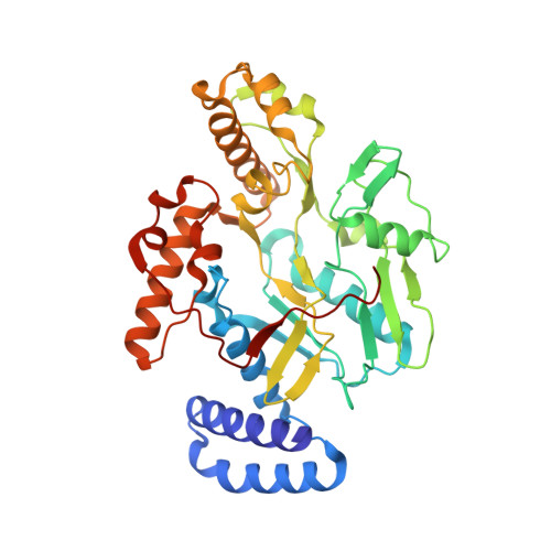
    Nitric oxide generated by bacterial nitric oxide synthase (NOS) increases the susceptibility of Gram-positive pathogens Staphylococcus aureus and Bacillus anthracis to oxidative stress, including antibiotic-induced oxidative stress. Not surprisingly, NOS inhibitors also improve the effectiveness of antimicrobials. Development of potent and selective bacterial NOS inhibitors is complicated by the high active site sequence and structural conservation shared with the mammalian NOS isoforms. To exploit bacterial NOS for the development of new therapeutics, recognition of alternative NOS surfaces and pharmacophores suitable for drug binding is required. Here, we report on a wide number of inhibitor-bound bacterial NOS crystal structures to identify several compounds that interact with surfaces unique to the bacterial NOS. Although binding studies indicate that these inhibitors weakly interact with the NOS active site, many of the inhibitors reported here provide a revised structural framework for the development of new antimicrobials that target bacterial NOS. In addition, mutagenesis studies reveal several key residues that unlock access to bacterial NOS surfaces that could provide the selectivity required to develop potent bacterial NOS inhibitors.


  • Organizational Affiliation
    • Departments of †Molecular Biology and Biochemistry, ‡Pharmaceutical Sciences, and §Chemistry, University of California, Irvine, California 92697-3900, United States.

Macromolecule Content 

  • Total Structure Weight: 43.18 kDa 
  • Atom Count: 3,320 
  • Modeled Residue Count: 362 
  • Deposited Residue Count: 363 
  • Unique protein chains: 1

Macromolecules

Find similar proteins by:|  3D Structure
Entity ID: 1
MoleculeChains  Sequence LengthOrganismDetailsImage
NITRIC OXIDE SYNTHASE OXYGENASE363Bacillus subtilis subsp. subtilis str. 168Mutation(s): 3 
EC: 1.14.13.165 (PDB Primary Data), 1.14.14.47 (UniProt)
UniProt
Find proteins for O34453 (Bacillus subtilis (strain 168))
Explore O34453 
Go to UniProtKB:  O34453
Entity Groups
Sequence Clusters30% Identity50% Identity70% Identity90% Identity95% Identity100% Identity
UniProt GroupO34453
Sequence Annotations
Expand
Reference Sequence

Small Molecules

Ligands 5 Unique
IDChains Name / Formula / InChI Key2D Diagram3D Interactions
HEM

Query on HEM


Download:Ideal Coordinates CCD File
B [auth A]PROTOPORPHYRIN IX CONTAINING FE
C34 H32 Fe N4 O4
KABFMIBPWCXCRK-RGGAHWMASA-L
H49

Query on H49


Download:Ideal Coordinates CCD File
E [auth A](S)-N-(3-(((Pyrrolidin-2-ylmethyl)amino)methyl)phenyl)thiophene-2-carboximidamide
C17 H22 N4 S
MRTCFHYZEYSLIF-HNNXBMFYSA-N
H4B

Query on H4B


Download:Ideal Coordinates CCD File
C [auth A]5,6,7,8-TETRAHYDROBIOPTERIN
C9 H15 N5 O3
FNKQXYHWGSIFBK-RPDRRWSUSA-N
GOL

Query on GOL


Download:Ideal Coordinates CCD File
F [auth A],
G [auth A]
GLYCEROL
C3 H8 O3
PEDCQBHIVMGVHV-UHFFFAOYSA-N
CL

Query on CL


Download:Ideal Coordinates CCD File
D [auth A]CHLORIDE ION
Cl
VEXZGXHMUGYJMC-UHFFFAOYSA-M
Binding Affinity Annotations 
IDSourceBinding Affinity
H49 BindingDB:  4UGN Kd: 1.78e+4 (nM) from 1 assay(s)

Experimental Data & Validation

Experimental Data

  • Method: X-RAY DIFFRACTION
  • Resolution: 2.09 Å
  • R-Value Free:  0.200 (Depositor), 0.199 (DCC) 
  • R-Value Work:  0.156 (Depositor), 0.156 (DCC) 
  • R-Value Observed: 0.158 (Depositor) 
Space Group: P 21 21 2
Unit Cell:
Length ( Å )Angle ( ˚ )
a = 80.65α = 90
b = 94.94β = 90
c = 62.92γ = 90
Software Package:
Software NamePurpose
PHENIXrefinement
MOSFLMdata reduction
Aimlessdata scaling

Structure Validation

View Full Validation Report


Ligand Structure Quality Assessment 


Entry History 

Deposition Data

Revision History  (Full details and data files)

  • Version 1.0: 2015-06-24
    Type: Initial release
  • Version 1.1: 2015-07-22
    Changes: Database references
  • Version 1.2: 2019-01-30
    Changes: Data collection, Experimental preparation, Other
  • Version 1.3: 2019-02-06
    Changes: Data collection, Experimental preparation
  • Version 1.4: 2023-12-20
    Changes: Data collection, Database references, Derived calculations, Other, Refinement description