4UDD | pdb_00004udd

GR in complex with desisobutyrylciclesonide


Experimental Data Snapshot

  • Method: X-RAY DIFFRACTION
  • Resolution: 1.80 Å
  • R-Value Free: 
    0.224 (Depositor), 0.206 (DCC) 
  • R-Value Work: 
    0.213 (Depositor), 0.192 (DCC) 
  • R-Value Observed: 
    0.213 (Depositor) 

Starting Model: experimental
View more details

wwPDB Validation 3D Report Full Report

Validation slider image for 4UDD

Ligand Structure Quality Assessment 


This is version 1.5 of the entry. See complete history

Literature

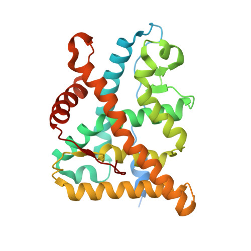
Ligand Binding Mechanism in Steroid Receptors: From Conserved Plasticity to Differential Evolutionary Constraints.

Edman, K.Hosseini, A.Bjursell, M.K.Aagaard, A.Wissler, L.Gunnarsson, A.Kaminski, T.Kohler, C.Backstrom, S.Jensen, T.J.Cavallin, A.Karlsson, U.Nilsson, E.Lecina, D.Takahashi, R.Grebner, C.Geschwindner, S.Lepisto, M.Hogner, A.C.Guallar, V.

(2015) Structure 23: 2280

  • DOI: https://doi.org/10.1016/j.str.2015.09.012
  • Primary Citation Related Structures: 
    4UDA, 4UDB, 4UDC, 4UDD

  • PubMed Abstract: 

    Steroid receptor drugs have been available for more than half a century, but details of the ligand binding mechanism have remained elusive. We solved X-ray structures of the glucocorticoid and mineralocorticoid receptors to identify a conserved plasticity at the helix 6-7 region that extends the ligand binding pocket toward the receptor surface. Since none of the endogenous ligands exploit this region, we hypothesized that it constitutes an integral part of the binding event. Extensive all-atom unbiased ligand exit and entrance simulations corroborate a ligand binding pathway that gives the observed structural plasticity a key functional role. Kinetic measurements reveal that the receptor residence time correlates with structural rearrangements observed in both structures and simulations. Ultimately, our findings reveal why nature has conserved the capacity to open up this region, and highlight how differences in the details of the ligand entry process result in differential evolutionary constraints across the steroid receptors.


  • Organizational Affiliation
    • Discovery Sciences, AstraZeneca, Mölndal, Pepparedsleden 1, 43183 Mölndal, Sweden. Electronic address: karl.edman@astrazeneca.com.

Macromolecule Content 

  • Total Structure Weight: 36.27 kDa 
  • Atom Count: 2,580 
  • Modeled Residue Count: 262 
  • Deposited Residue Count: 294 
  • Unique protein chains: 2

Macromolecules

Find similar proteins by:|  3D Structure
Entity ID: 1
MoleculeChains  Sequence LengthOrganismDetailsImage
GLUCOCORTICOID RECEPTOR280Homo sapiensMutation(s): 4 
UniProt & NIH Common Fund Data Resources
Find proteins for P04150 (Homo sapiens)
Explore P04150 
Go to UniProtKB:  P04150
PHAROS:  P04150
GTEx:  ENSG00000113580 
Entity Groups
Sequence Clusters30% Identity50% Identity70% Identity90% Identity95% Identity100% Identity
UniProt GroupP04150
Sequence Annotations
Expand
Reference Sequence
Find similar proteins by:  Sequence   |   3D Structure  
Entity ID: 2
MoleculeChains  Sequence LengthOrganismDetailsImage
NUCLEAR RECEPTOR COACTIVATOR 214Homo sapiensMutation(s): 0 
UniProt & NIH Common Fund Data Resources
Find proteins for Q15596 (Homo sapiens)
Explore Q15596 
Go to UniProtKB:  Q15596
PHAROS:  Q15596
GTEx:  ENSG00000140396 
Entity Groups
Sequence Clusters30% Identity50% Identity70% Identity90% Identity95% Identity100% Identity
UniProt GroupQ15596
Sequence Annotations
Expand
Reference Sequence

Small Molecules

Experimental Data & Validation

Experimental Data

  • Method: X-RAY DIFFRACTION
  • Resolution: 1.80 Å
  • R-Value Free:  0.224 (Depositor), 0.206 (DCC) 
  • R-Value Work:  0.213 (Depositor), 0.192 (DCC) 
  • R-Value Observed: 0.213 (Depositor) 
Space Group: P 32 2 1
Unit Cell:
Length ( Å )Angle ( ˚ )
a = 87.2α = 90
b = 87.2β = 90
c = 102.887γ = 120
Software Package:
Software NamePurpose
BUSTERrefinement
MOSFLMdata reduction
SCALAdata scaling
PHASERphasing

Structure Validation

View Full Validation Report


Ligand Structure Quality Assessment 


Entry History 

Revision History  (Full details and data files)

  • Version 1.0: 2015-11-25
    Type: Initial release
  • Version 1.1: 2015-12-09
    Changes: Database references
  • Version 1.2: 2015-12-16
    Changes: Database references
  • Version 1.3: 2017-09-27
    Changes: Data collection
  • Version 1.4: 2019-04-03
    Changes: Data collection, Source and taxonomy
  • Version 1.5: 2023-12-20
    Changes: Data collection, Database references, Derived calculations, Other, Refinement description