4U7M | pdb_00004u7m

LRIG1 extracellular domain: Structure and Function Analysis

  • Classification: SIGNALING PROTEIN
  • Organism(s): Homo sapiens
  • Expression System: Spodoptera frugiperda
  • Mutation(s): No 

  • Deposited: 2014-07-31 Released: 2015-04-08 
  • Deposition Author(s): Xu, Y.
  • Funding Organization(s): National Health and Medical Research Council (NHMRC, Australia)

Experimental Data Snapshot

  • Method: X-RAY DIFFRACTION
  • Resolution: 2.76 Å
  • R-Value Free: 
    0.253 (Depositor), 0.255 (DCC) 
  • R-Value Work: 
    0.205 (Depositor), 0.213 (DCC) 
  • R-Value Observed: 
    0.207 (Depositor) 

Starting Model: experimental
View more details

wwPDB Validation 3D Report Full Report

Validation slider image for 4U7M

Ligand Structure Quality Assessment 


This is version 1.6 of the entry. See complete history

Literature

LRIG1 Extracellular Domain: Structure and Function Analysis.

Xu, Y.Soo, P.Walker, F.Zhang, H.H.Redpath, N.Tan, C.W.Nicola, N.A.Adams, T.E.Garrett, T.P.Zhang, J.G.Burgess, A.W.

(2015) J Mol Biology 427: 1934-1948

  • DOI: https://doi.org/10.1016/j.jmb.2015.03.001
  • Primary Citation Related Structures: 
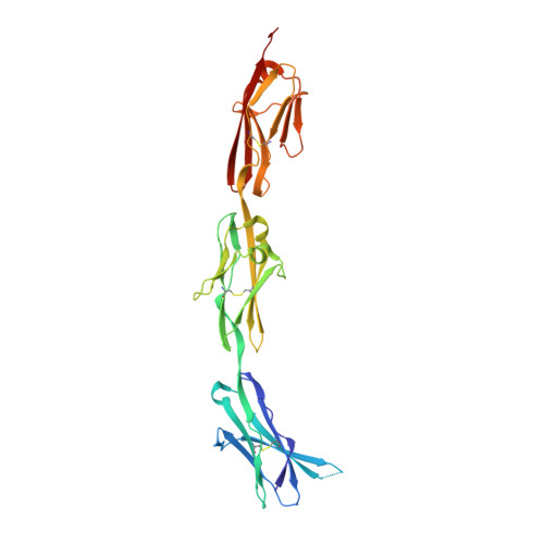
    4U7L, 4U7M

  • PubMed Abstract: 

    We have expressed and purified three soluble fragments of the human LRIG1-ECD (extracellular domain): the LRIG1-LRR (leucine-rich repeat) domain, the LRIG1-3Ig (immunoglobulin-like) domain, and the LRIG1-LRR-1Ig fragment using baculovirus vectors in insect cells. The two LRIG1 domains crystallised so that we have been able to determine the three-dimensional structures at 2.3Å resolution. We developed a three-dimensional structure for the LRIG1-ECD using homology modelling based on the LINGO-1 structure. The LRIG1-LRR domain and the LRIG1-LRR-1Ig fragment are monomers in solution, whereas the LRIG1-3Ig domain appears to be dimeric. We could not detect any binding of the LRIG1 domains or the LRIG1-LRR-1Ig fragment to the EGF receptor (EGFR), either in solution using biosensor analysis or when the EGFR was expressed on the cell surface. The FLAG-tagged LRIG1-LRR-1Ig fragment binds weakly to colon cancer cells regardless of the presence of EGFRs. Similarly, neither the soluble LRIG1-LRR nor the LRIG1-3Ig domains nor the full-length LRIG1 co-expressed in HEK293 cells inhibited ligand-stimulated activation of cell-surface EGFR.


  • Organizational Affiliation
    • Structural Biology Division, The Walter and Eliza Hall Institute of Medical Research, Parkville, Victoria 3052, Australia; Cancer and Haematology Division, The Walter and Eliza Hall Institute of Medical Research, Parkville, Victoria 3052, Australia.

Macromolecule Content 

  • Total Structure Weight: 32.65 kDa 
  • Atom Count: 2,264 
  • Modeled Residue Count: 289 
  • Deposited Residue Count: 293 
  • Unique protein chains: 1

Macromolecules

Find similar proteins by:|  3D Structure
Entity ID: 1
MoleculeChains  Sequence LengthOrganismDetailsImage
Leucine-rich repeats and immunoglobulin-like domains protein 1293Homo sapiensMutation(s): 0 
Gene Names: LRIG1LIG1
UniProt & NIH Common Fund Data Resources
Find proteins for Q96JA1 (Homo sapiens)
Explore Q96JA1 
Go to UniProtKB:  Q96JA1
PHAROS:  Q96JA1
GTEx:  ENSG00000144749 
Entity Groups
Sequence Clusters30% Identity50% Identity70% Identity90% Identity95% Identity100% Identity
UniProt GroupQ96JA1
Glycosylation
Glycosylation Sites: 1Go to GlyGen: Q96JA1-1
Sequence Annotations
Expand
Reference Sequence

Small Molecules

Ligands 1 Unique
IDChains Name / Formula / InChI Key2D Diagram3D Interactions
NAG

Query on NAG


Download:Ideal Coordinates CCD File
B [auth A]2-acetamido-2-deoxy-beta-D-glucopyranose
C8 H15 N O6
OVRNDRQMDRJTHS-FMDGEEDCSA-N

Experimental Data & Validation

Experimental Data

  • Method: X-RAY DIFFRACTION
  • Resolution: 2.76 Å
  • R-Value Free:  0.253 (Depositor), 0.255 (DCC) 
  • R-Value Work:  0.205 (Depositor), 0.213 (DCC) 
  • R-Value Observed: 0.207 (Depositor) 
Space Group: P 43 21 2
Unit Cell:
Length ( Å )Angle ( ˚ )
a = 121.04α = 90
b = 121.04β = 90
c = 115.8γ = 90
Software Package:
Software NamePurpose
PHENIXrefinement
XDSdata reduction
PDB_EXTRACTdata extraction
XSCALEdata scaling
PHASERphasing

Structure Validation

View Full Validation Report


Ligand Structure Quality Assessment 


Entry History 

& Funding Information

Deposition Data

  • Released Date: 2015-04-08 
  • Deposition Author(s): Xu, Y.

Funding OrganizationLocationGrant Number
National Health and Medical Research Council (NHMRC, Australia)Australia487922

Revision History  (Full details and data files)

  • Version 1.0: 2015-04-08
    Type: Initial release
  • Version 1.1: 2015-05-13
    Changes: Database references
  • Version 1.2: 2017-09-20
    Changes: Author supporting evidence, Data collection, Database references, Derived calculations, Source and taxonomy
  • Version 1.3: 2020-01-08
    Changes: Author supporting evidence
  • Version 1.4: 2020-07-29
    Type: Remediation
    Reason: Carbohydrate remediation
    Changes: Data collection, Derived calculations, Structure summary
  • Version 1.5: 2023-09-27
    Changes: Data collection, Database references, Refinement description, Structure summary
  • Version 1.6: 2024-10-23
    Changes: Structure summary