4U5D | pdb_00004u5d

Crystal structure of GluA2, con-ikot-ikot snail toxin, partial agonist KA and postitive modulator (R,R)-2b complex


Experimental Data Snapshot

  • Method: X-RAY DIFFRACTION
  • Resolution: 3.58 Å
  • R-Value Free: 
    0.297 (Depositor), 0.302 (DCC) 
  • R-Value Work: 
    0.256 (Depositor), 0.265 (DCC) 
  • R-Value Observed: 
    0.258 (Depositor) 

wwPDB Validation 3D Report Full Report

Validation slider image for 4U5D

Ligand Structure Quality Assessment 


This is version 1.6 of the entry. See complete history

Literature

X-ray structures of AMPA receptor-cone snail toxin complexes illuminate activation mechanism.

Chen, L.Durr, K.L.Gouaux, E.

(2014) Science 345: 1021-1026

  • DOI: https://doi.org/10.1126/science.1258409
  • Primary Citation Related Structures: 
    4U5B, 4U5C, 4U5D, 4U5E, 4U5F, 4U5G, 4U5H

  • PubMed Abstract: 

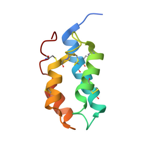
    AMPA-sensitive glutamate receptors are crucial to the structural and dynamic properties of the brain, to the development and function of the central nervous system, and to the treatment of neurological conditions from depression to cognitive impairment. However, the molecular principles underlying AMPA receptor activation have remained elusive. We determined multiple x-ray crystal structures of the GluA2 AMPA receptor in complex with a Conus striatus cone snail toxin, a positive allosteric modulator, and orthosteric agonists, at 3.8 to 4.1 angstrom resolution. We show how the toxin acts like a straightjacket on the ligand-binding domain (LBD) "gating ring," restraining the domains via both intra- and interdimer cross-links such that agonist-induced closure of the LBD "clamshells" is transduced into an irislike expansion of the gating ring. By structural analysis of activation-enhancing mutants, we show how the expansion of the LBD gating ring results in pulling forces on the M3 helices that, in turn, are coupled to ion channel gating.


  • Organizational Affiliation
    • Vollum Institute, Oregon Health and Science University, 3181 SW Sam Jackson Park Road, Portland, OR 97239, USA.

Macromolecule Content 

  • Total Structure Weight: 387.75 kDa 
  • Atom Count: 23,720 
  • Modeled Residue Count: 3,132 
  • Deposited Residue Count: 3,436 
  • Unique protein chains: 2

Macromolecules

Find similar proteins by:|  3D Structure
Entity ID: 1
MoleculeChains  Sequence LengthOrganismDetailsImage
Glutamate receptor 2
A, B, C, D
814Rattus norvegicusMutation(s): 23 
Gene Names: Gria2Glur2
Membrane Entity: Yes 
UniProt
Find proteins for P19491 (Rattus norvegicus)
Explore P19491 
Go to UniProtKB:  P19491
Entity Groups
Sequence Clusters30% Identity50% Identity70% Identity90% Identity95% Identity100% Identity
UniProt GroupP19491
Glycosylation
Glycosylation Sites: 1Go to GlyGen: P19491-1
Sequence Annotations
Expand
Reference Sequence
Find similar proteins by:|  3D Structure
Entity ID: 2
MoleculeChains  Sequence LengthOrganismDetailsImage
Con-ikot-ikot
E, F
90Conus striatusMutation(s): 0 
Membrane Entity: Yes 
UniProt
Find proteins for P0CB20 (Conus striatus)
Explore P0CB20 
Go to UniProtKB:  P0CB20
Entity Groups
Sequence Clusters30% Identity50% Identity70% Identity90% Identity95% Identity100% Identity
UniProt GroupP0CB20
Sequence Annotations
Expand
Reference Sequence

Small Molecules

Ligands 3 Unique
IDChains Name / Formula / InChI Key2D Diagram3D Interactions
FWF

Query on FWF


Download:Ideal Coordinates CCD File
I [auth A],
L [auth B]
N,N'-[biphenyl-4,4'-diyldi(2R)propane-2,1-diyl]dipropane-2-sulfonamide
C24 H36 N2 O4 S2
HGLQSTHVRKGLQP-PMACEKPBSA-N
NAG

Query on NAG


Download:Ideal Coordinates CCD File
H [auth A],
K [auth B],
N [auth C],
P [auth D]
2-acetamido-2-deoxy-beta-D-glucopyranose
C8 H15 N O6
OVRNDRQMDRJTHS-FMDGEEDCSA-N
KAI

Query on KAI


Download:Ideal Coordinates CCD File
G [auth A],
J [auth B],
M [auth C],
O [auth D]
3-(CARBOXYMETHYL)-4-ISOPROPENYLPROLINE
C10 H15 N O4
VLSMHEGGTFMBBZ-OOZYFLPDSA-N

Experimental Data & Validation

Experimental Data

  • Method: X-RAY DIFFRACTION
  • Resolution: 3.58 Å
  • R-Value Free:  0.297 (Depositor), 0.302 (DCC) 
  • R-Value Work:  0.256 (Depositor), 0.265 (DCC) 
  • R-Value Observed: 0.258 (Depositor) 
Space Group: P 21 21 2
Unit Cell:
Length ( Å )Angle ( ˚ )
a = 161.51α = 90
b = 367.58β = 90
c = 109.38γ = 90
Software Package:
Software NamePurpose
PHENIXrefinement
XDSdata scaling
PDB_EXTRACTdata extraction

Structure Validation

View Full Validation Report


Ligand Structure Quality Assessment 


Entry History 

Deposition Data

Revision History  (Full details and data files)

  • Version 1.0: 2014-08-13
    Type: Initial release
  • Version 1.1: 2014-08-27
    Changes: Database references
  • Version 1.2: 2014-10-01
    Changes: Database references
  • Version 1.3: 2017-11-22
    Changes: Database references, Derived calculations, Other, Refinement description, Source and taxonomy, Structure summary
  • Version 1.4: 2020-07-29
    Type: Remediation
    Reason: Carbohydrate remediation
    Changes: Data collection, Derived calculations, Refinement description, Structure summary
  • Version 1.5: 2023-12-27
    Changes: Data collection, Database references, Structure summary
  • Version 1.6: 2024-10-23
    Changes: Structure summary