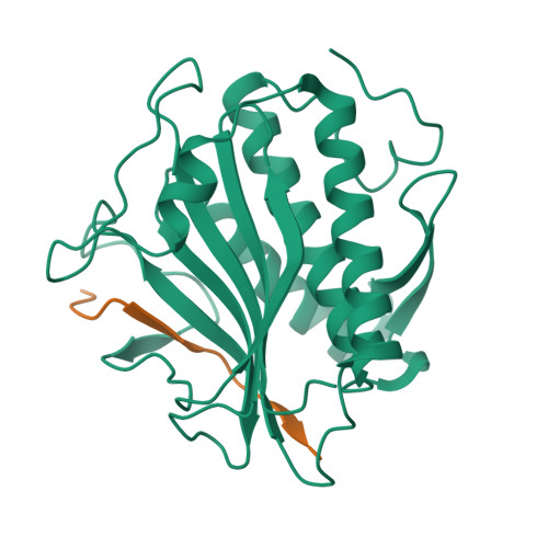
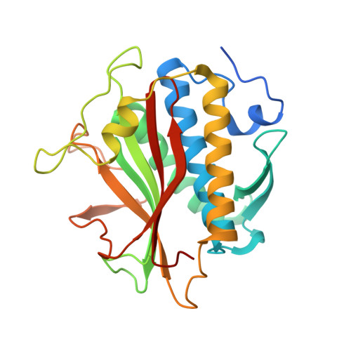
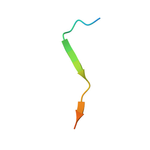
4TZO

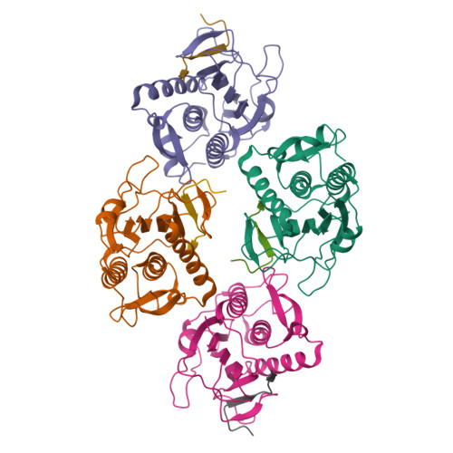
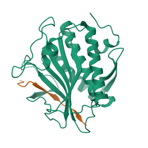
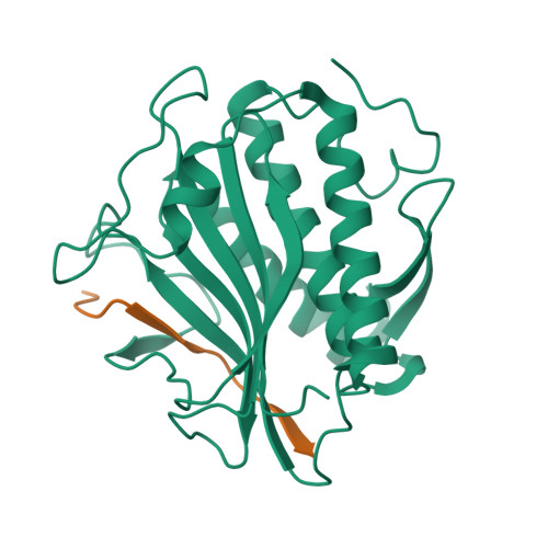
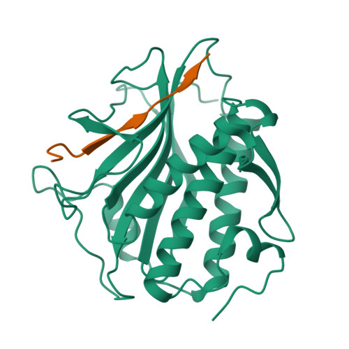
Structure of C. elegans HTP-1 bound to HIM-3 closure motif


Experimental Data Snapshot

  • Method: X-RAY DIFFRACTION
  • Resolution: 2.40 Å
  • R-Value Free: 0.282 
  • R-Value Work: 0.246 
  • R-Value Observed: 0.247 

Starting Model: experimental
View more details

wwPDB Validation   3D Report Full Report


This is version 1.4 of the entry. See complete history


Literature

The Chromosome Axis Controls Meiotic Events through a Hierarchical Assembly of HORMA Domain Proteins.

Kim, Y.Rosenberg, S.C.Kugel, C.L.Kostow, N.Rog, O.Davydov, V.Su, T.Y.Dernburg, A.F.Corbett, K.D.

(2014) Dev Cell 31: 487-502

  • DOI: https://doi.org/10.1016/j.devcel.2014.09.013
  • Primary Citation of Related Structures:  
    4TRK, 4TZJ, 4TZL, 4TZM, 4TZN, 4TZO, 4TZQ, 4TZS

  • PubMed Abstract: 

    Proteins of the HORMA domain family play central, but poorly understood, roles in chromosome organization and dynamics during meiosis. In Caenorhabditis elegans, four such proteins (HIM-3, HTP-1, HTP-2, and HTP-3) have distinct but overlapping functions. Through combined biochemical, structural, and in vivo analysis, we find that these proteins form hierarchical complexes through binding of their HORMA domains to cognate peptides within their partners' C-terminal tails, analogous to the "safety belt" binding mechanism of Mad2. These interactions are critical for recruitment of HIM-3, HTP-1, and HTP-2 to chromosome axes. HTP-3, in addition to recruiting the other HORMA domain proteins to the axis, plays an independent role in sister chromatid cohesion and double-strand break formation. Finally, we find that mammalian HORMAD1 binds a motif found both at its own C terminus and at that of HORMAD2, indicating that this mode of intermolecular association is a conserved feature of meiotic chromosome structure in eukaryotes.


  • Organizational Affiliation

    Department of Molecular and Cell Biology, University of California, Berkeley, Berkeley, CA 94720-3220, USA; Howard Hughes Medical Institute, 4000 Jones Bridge Road, Chevy Chase, MD 20815, USA; Life Sciences Division, Department of Genome Dynamics, Lawrence Berkeley National Laboratory, Berkeley, CA 94720, USA; California Institute for Quantitative Biosciences, Berkeley, CA 94720, USA.


Macromolecules
Find similar proteins by:  (by identity cutoff)  |  3D Structure
Entity ID: 1
MoleculeChains Sequence LengthOrganismDetailsImage
Protein HTP-1
A, C, E, G
253Caenorhabditis elegansMutation(s): 1 
Gene Names: htp-1CELE_F41H10.10F41H10.10
UniProt
Find proteins for Q20305 (Caenorhabditis elegans)
Explore Q20305 
Go to UniProtKB:  Q20305
Entity Groups  
Sequence Clusters30% Identity50% Identity70% Identity90% Identity95% Identity100% Identity
UniProt GroupQ20305
Sequence Annotations
Expand
  • Reference Sequence

Find similar proteins by:  Sequence   |   3D Structure  

Entity ID: 2
MoleculeChains Sequence LengthOrganismDetailsImage
C. elegans HIM-3 closure motif
B, D, F, H
20Caenorhabditis elegansMutation(s): 0 
Gene Names: him-3CELE_ZK381.1ZK381.1
UniProt
Find proteins for G5EBG0 (Caenorhabditis elegans)
Explore G5EBG0 
Go to UniProtKB:  G5EBG0
Entity Groups  
Sequence Clusters30% Identity50% Identity70% Identity90% Identity95% Identity100% Identity
UniProt GroupG5EBG0
Sequence Annotations
Expand
  • Reference Sequence
Experimental Data & Validation

Experimental Data

Unit Cell:
Length ( Å )Angle ( ˚ )
a = 66.897α = 90
b = 107.873β = 90.1
c = 82.968γ = 90
Software Package:
Software NamePurpose
PHENIXrefinement

Structure Validation

View Full Validation Report


Entry History & Funding Information

Deposition Data


Funding OrganizationLocationGrant Number
National Institutes of Health/National Institute of General Medical Sciences (NIH/NIGMS)United StatesR01GM104141

Revision History  (Full details and data files)

  • Version 1.0: 2014-11-19
    Type: Initial release
  • Version 1.1: 2014-12-17
    Changes: Database references
  • Version 1.2: 2017-09-20
    Changes: Author supporting evidence, Derived calculations, Source and taxonomy, Structure summary
  • Version 1.3: 2019-12-25
    Changes: Author supporting evidence
  • Version 1.4: 2023-09-27
    Changes: Data collection, Database references, Refinement description