4TZA | pdb_00004tza

TGP, an extremely thermostable green fluorescent protein created by structure-guided surface engineering


Experimental Data Snapshot

  • Method: X-RAY DIFFRACTION
  • Resolution: 1.90 Å
  • R-Value Free: 
    0.200 (Depositor) 
  • R-Value Work: 
    0.174 (Depositor) 
  • R-Value Observed: 
    0.175 (Depositor) 

wwPDB Validation 3D Report Full Report

Validation slider image for 4TZA

This is version 2.1 of the entry. See complete history

Literature

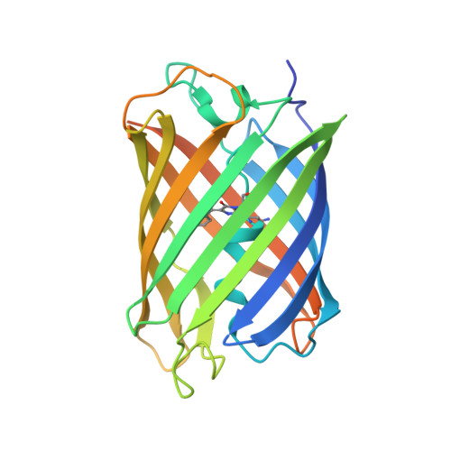
Thermal green protein, an extremely stable, nonaggregating fluorescent protein created by structure-guided surface engineering.

Close, D.W.Paul, C.D.Langan, P.S.Wilce, M.C.Traore, D.A.Halfmann, R.Rocha, R.C.Waldo, G.S.Payne, R.J.Rucker, J.B.Prescott, M.Bradbury, A.R.

(2015) Proteins 83: 1225-1237

  • DOI: https://doi.org/10.1002/prot.24699
  • Primary Citation Related Structures: 
    4TZA, 4TZG

  • PubMed Abstract: 

    In this article, we describe the engineering and X-ray crystal structure of Thermal Green Protein (TGP), an extremely stable, highly soluble, non-aggregating green fluorescent protein. TGP is a soluble variant of the fluorescent protein eCGP123, which despite being highly stable, has proven to be aggregation-prone. The X-ray crystal structure of eCGP123, also determined within the context of this paper, was used to carry out rational surface engineering to improve its solubility, leading to TGP. The approach involved simultaneously eliminating crystal lattice contacts while increasing the overall negative charge of the protein. Despite intentional disruption of lattice contacts and introduction of high entropy glutamate side chains, TGP crystallized readily in a number of different conditions and the X-ray crystal structure of TGP was determined to 1.9 Å resolution. The structural reasons for the enhanced stability of TGP and eCGP123 are discussed. We demonstrate the utility of using TGP as a fusion partner in various assays and significantly, in amyloid assays in which the standard fluorescent protein, EGFP, is undesirable because of aberrant oligomerization.


  • Organizational Affiliation
    • Bioscience Division, Los Alamos National Laboratory, Los Alamos, New Mexico.

Macromolecule Content 

  • Total Structure Weight: 113.01 kDa 
  • Atom Count: 7,832 
  • Modeled Residue Count: 858 
  • Deposited Residue Count: 996 
  • Unique protein chains: 1

Macromolecules

Find similar proteins by:|  3D Structure
Entity ID: 1
MoleculeChains  Sequence LengthOrganismDetailsImage
Fluorescent ProteinA [auth C],
B [auth A],
C [auth B],
D
249synthetic constructMutation(s): 0 

Small Molecules

Modified Residues  1 Unique
IDChains TypeFormula2D DiagramParent
CRQ
Query on CRQ
A [auth C],
B [auth A],
C [auth B],
D
L-PEPTIDE LINKINGC16 H16 N4 O5GLN, TYR, GLY

Experimental Data & Validation

Experimental Data

  • Method: X-RAY DIFFRACTION
  • Resolution: 1.90 Å
  • R-Value Free:  0.200 (Depositor) 
  • R-Value Work:  0.174 (Depositor) 
  • R-Value Observed: 0.175 (Depositor) 
Space Group: P 21 21 21
Unit Cell:
Length ( Å )Angle ( ˚ )
a = 94.1999α = 90
b = 141.22β = 90
c = 69.0897γ = 90
Software Package:
Software NamePurpose
HKL-3000data collection
iMOSFLMdata reduction
SCALAdata scaling
Cootmodel building
PHENIXphasing
PHENIXrefinement

Structure Validation

View Full Validation Report


Entry History 

Deposition Data

Revision History  (Full details and data files)

  • Version 1.0: 2014-10-22
    Type: Initial release
  • Version 1.1: 2014-10-29
    Changes: Database references
  • Version 1.2: 2015-02-04
    Changes: Derived calculations
  • Version 1.3: 2015-06-03
    Changes: Database references
  • Version 1.4: 2016-02-17
    Changes: Data collection, Database references
  • Version 2.0: 2023-11-15
    Changes: Advisory, Atomic model, Data collection, Database references, Derived calculations
  • Version 2.1: 2024-10-16
    Changes: Structure summary