4TU7 | pdb_00004tu7

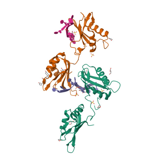
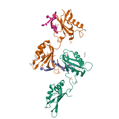
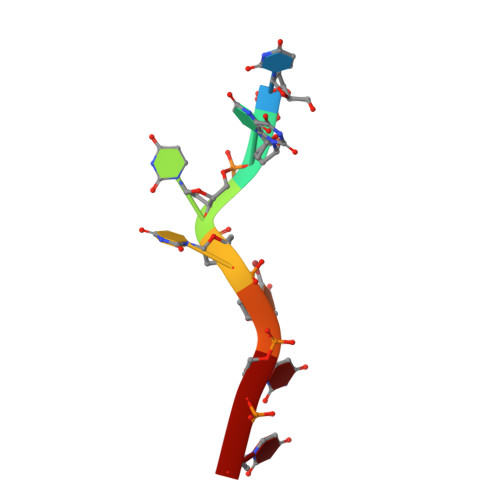
Structure of U2AF65 D231V variant with BrU5 DNA


Experimental Data Snapshot

  • Method: X-RAY DIFFRACTION
  • Resolution: 2.09 Å
  • R-Value Free: 
    0.223 (Depositor), 0.230 (DCC) 
  • R-Value Work: 
    0.174 (Depositor), 0.180 (DCC) 
  • R-Value Observed: 
    0.179 (Depositor) 

wwPDB Validation   3D Report Full Report


Ligand Structure Quality Assessment 

Created with Raphaël 2.3.0Worse 01 BetterLigand structure goodness of fit to experimental dataBest fitted CPQClick on this verticalbar to view details

This is version 1.4 of the entry. See complete history


Literature

Structure-guided U2AF65 variant improves recognition and splicing of a defective pre-mRNA.

Agrawal, A.A.McLaughlin, K.J.Jenkins, J.L.Kielkopf, C.L.

(2014) Proc Natl Acad Sci U S A 111: 17420-17425

  • DOI: https://doi.org/10.1073/pnas.1412743111
  • Primary Citation of Related Structures:  
    4TU7, 4TU8, 4TU9

  • PubMed Abstract: 

    Purine interruptions of polypyrimidine (Py) tract splice site signals contribute to human genetic diseases. The essential splicing factor U2AF(65) normally recognizes a Py tract consensus sequence preceding the major class of 3' splice sites. We found that neurofibromatosis- or retinitis pigmentosa-causing mutations in the 5' regions of Py tracts severely reduce U2AF(65) affinity. Conversely, we identified a preferred binding site of U2AF(65) for purine substitutions in the 3' regions of Py tracts. Based on a comparison of new U2AF(65) structures bound to either A- or G-containing Py tracts with previously identified pyrimidine-containing structures, we expected to find that a D231V amino acid change in U2AF(65) would specify U over other nucleotides. We found that the crystal structure of the U2AF(65)-D231V variant confirms favorable packing between the engineered valine and a target uracil base. The D231V amino acid change restores U2AF(65) affinity for two mutated splice sites that cause human genetic diseases and successfully promotes splicing of a defective retinitis pigmentosa-causing transcript. We conclude that reduced U2AF(65) binding is a molecular consequence of disease-relevant mutations, and that a structure-guided U2AF(65) variant is capable of manipulating gene expression in eukaryotic cells.


  • Organizational Affiliation

    Center for RNA Biology and Department of Biochemistry and Biophysics, University of Rochester School of Medicine and Dentistry, Rochester, NY 14642.


Macromolecules

Find similar proteins by:  (by identity cutoff)  |  3D Structure
Entity ID: 1
MoleculeChains Sequence LengthOrganismDetailsImage
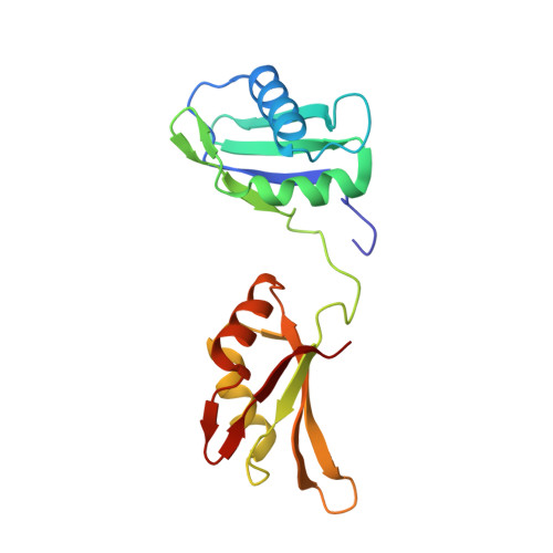
Splicing factor U2AF 65 kDa subunit
A, B
174Homo sapiensMutation(s): 1 
Gene Names: U2AF2U2AF65
UniProt & NIH Common Fund Data Resources
Find proteins for P26368 (Homo sapiens)
Explore P26368 
Go to UniProtKB:  P26368
PHAROS:  P26368
GTEx:  ENSG00000063244 
Entity Groups  
Sequence Clusters30% Identity50% Identity70% Identity90% Identity95% Identity100% Identity
UniProt GroupP26368
Sequence Annotations
Expand
  • Reference Sequence
Small Molecules
Ligands 4 Unique
IDChains Name / Formula / InChI Key2D Diagram3D Interactions
CPQ
Query on CPQ

Download Ideal Coordinates CCD File 
O [auth B]N,N-BIS(3-D-GLUCONAMIDOPROPYL)DEOXYCHOLAMIDE
C42 H75 N3 O15
OJSUWTDDXLCUFR-HGZMBBKESA-N
SO4
Query on SO4

Download Ideal Coordinates CCD File 
G [auth A]
H [auth A]
I [auth A]
P [auth B]
Q [auth B]
SULFATE ION
O4 S
QAOWNCQODCNURD-UHFFFAOYSA-L
GOL
Query on GOL

Download Ideal Coordinates CCD File 
J [auth A],
K [auth A],
L [auth A],
S [auth B]
GLYCEROL
C3 H8 O3
PEDCQBHIVMGVHV-UHFFFAOYSA-N
DIO
Query on DIO

Download Ideal Coordinates CCD File 
E [auth A],
F [auth A],
M [auth B],
N [auth B]
1,4-DIETHYLENE DIOXIDE
C4 H8 O2
RYHBNJHYFVUHQT-UHFFFAOYSA-N
Experimental Data & Validation

Experimental Data

  • Method: X-RAY DIFFRACTION
  • Resolution: 2.09 Å
  • R-Value Free:  0.223 (Depositor), 0.230 (DCC) 
  • R-Value Work:  0.174 (Depositor), 0.180 (DCC) 
  • R-Value Observed: 0.179 (Depositor) 
Space Group: C 1 2 1
Unit Cell:
Length ( Å )Angle ( ˚ )
a = 164.432α = 90
b = 37.361β = 125.6
c = 100.192γ = 90
Software Package:
Software NamePurpose
SCALAdata scaling
PHENIXrefinement
PDB_EXTRACTdata extraction

Structure Validation

View Full Validation Report


Ligand Structure Quality Assessment 

Created with Raphaël 2.3.0Worse 01 BetterLigand structure goodness of fit to experimental dataBest fitted CPQClick on this verticalbar to view details

Entry History 

Deposition Data

Revision History  (Full details and data files)

  • Version 1.0: 2014-11-26
    Type: Initial release
  • Version 1.1: 2014-12-10
    Changes: Database references
  • Version 1.2: 2014-12-24
    Changes: Database references
  • Version 1.3: 2017-11-22
    Changes: Database references, Derived calculations, Refinement description, Source and taxonomy
  • Version 1.4: 2023-12-27
    Changes: Data collection, Database references