4TU1 | pdb_00004tu1

Structure of Toxoplasma gondii fructose 1,6 bisphosphate aldolase


Experimental Data Snapshot

  • Method: X-RAY DIFFRACTION
  • Resolution: 2.00 Å
  • R-Value Free: 
    0.244 (Depositor), 0.246 (DCC) 
  • R-Value Work: 
    0.194 (Depositor), 0.198 (DCC) 
  • R-Value Observed: 
    0.196 (Depositor) 

Starting Model: experimental
View more details

wwPDB Validation 3D Report Full Report

Validation slider image for 4TU1

This is version 1.3 of the entry. See complete history

Literature

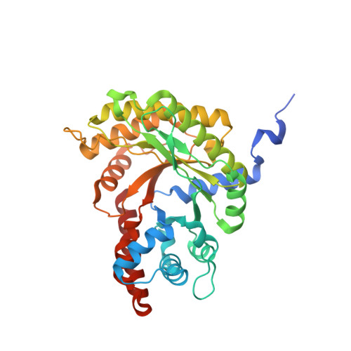
Structure of Toxoplasma gondii fructose-1,6-bisphosphate aldolase.

Boucher, L.E.Bosch, J.

(2014) Acta Crystallogr F Struct Biol Commun 70: 1186-1192

  • DOI: https://doi.org/10.1107/S2053230X14017087
  • Primary Citation Related Structures: 
    4TU1

  • PubMed Abstract: 

    The apicomplexan parasite Toxoplasma gondii must invade host cells to continue its lifecycle. It invades different cell types using an actomyosin motor that is connected to extracellular adhesins via the bridging protein fructose-1,6-bisphosphate aldolase. During invasion, aldolase serves in the role of a structural bridging protein, as opposed to its normal enzymatic role in the glycolysis pathway. Crystal structures of the homologous Plasmodium falciparum fructose-1,6-bisphosphate aldolase have been described previously. Here, T. gondii fructose-1,6-bisphosphate aldolase has been crystallized in space group P22121, with the biologically relevant tetramer in the asymmetric unit, and the structure has been determined via molecular replacement to a resolution of 2.0 Å. An analysis of the quality of the model and of the differences between the four chains in the asymmetric unit and a comparison between the T. gondii and P. falciparum aldolase structures is presented.


  • Organizational Affiliation
    • Department of Biochemistry and Molecular Biology, Johns Hopkins Bloomberg School of Public Health, 615 North Wolfe Street, Baltimore, MD 21205, USA.

Macromolecule Content 

  • Total Structure Weight: 154.31 kDa 
  • Atom Count: 11,268 
  • Modeled Residue Count: 1,354 
  • Deposited Residue Count: 1,420 
  • Unique protein chains: 1

Macromolecules

Find similar proteins by:|  3D Structure
Entity ID: 1
MoleculeChains  Sequence LengthOrganismDetailsImage
Fructose-1,6-bisphosphate aldolase
A, B, C, D
355Toxoplasma gondiiMutation(s): 0 
Gene Names: TGVEG_236040
EC: 4.1.2.13
UniProt
Find proteins for Q8I8I2 (Toxoplasma gondii)
Explore Q8I8I2 
Go to UniProtKB:  Q8I8I2
Entity Groups
Sequence Clusters30% Identity50% Identity70% Identity90% Identity95% Identity100% Identity
UniProt GroupQ8I8I2
Sequence Annotations
Expand
Reference Sequence

Small Molecules

Ligands 1 Unique
IDChains Name / Formula / InChI Key2D Diagram3D Interactions
GOL

Query on GOL


Download:Ideal Coordinates CCD File
E [auth A]GLYCEROL
C3 H8 O3
PEDCQBHIVMGVHV-UHFFFAOYSA-N

Experimental Data & Validation

Experimental Data

  • Method: X-RAY DIFFRACTION
  • Resolution: 2.00 Å
  • R-Value Free:  0.244 (Depositor), 0.246 (DCC) 
  • R-Value Work:  0.194 (Depositor), 0.198 (DCC) 
  • R-Value Observed: 0.196 (Depositor) 
Space Group: P 2 21 21
Unit Cell:
Length ( Å )Angle ( ˚ )
a = 92.258α = 90
b = 134.458β = 90
c = 162.43γ = 90
Software Package:
Software NamePurpose
PHENIXrefinement
Cootmodel building
Blu-Icedata collection
PHASERphasing
XDSdata scaling

Structure Validation

View Full Validation Report


Entry History 

Deposition Data

Revision History  (Full details and data files)

  • Version 1.0: 2014-09-03
    Type: Initial release
  • Version 1.1: 2014-09-24
    Changes: Database references
  • Version 1.2: 2017-11-22
    Changes: Derived calculations, Other, Refinement description, Source and taxonomy
  • Version 1.3: 2023-09-27
    Changes: Data collection, Database references, Refinement description