4TTN | pdb_00004ttn

Quasi-racemic structure of [G6A]kalata B1


Experimental Data Snapshot

  • Method: X-RAY DIFFRACTION
  • Resolution: 1.25 Å
  • R-Value Free: 
    0.229 (Depositor), 0.229 (DCC) 
  • R-Value Work: 
    0.212 (Depositor), 0.212 (DCC) 
  • R-Value Observed: 
    0.213 (Depositor) 

wwPDB Validation 3D Report Full Report

Validation slider image for 4TTN

This is version 1.4 of the entry. See complete history

Literature

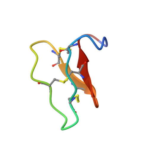
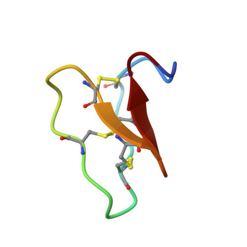
Racemic and Quasi-Racemic X-ray Structures of Cyclic Disulfide-Rich Peptide Drug Scaffolds.

Wang, C.K.King, G.J.Northfield, S.E.Ojeda, P.G.Craik, D.J.

(2014) Angew Chem Int Ed Engl 53: 11236-11241

  • DOI: https://doi.org/10.1002/anie.201406563
  • Primary Citation Related Structures: 
    4TTK, 4TTL, 4TTM, 4TTN, 4TTO

  • PubMed Abstract: 

    Cyclic disulfide-rich peptides have exceptional stability and are promising frameworks for drug design. We were interested in obtaining X-ray structures of these peptides to assist in drug design applications, but disulfide-rich peptides can be notoriously difficult to crystallize. To overcome this limitation, we chemically synthesized the L- and D-forms of three prototypic cyclic disulfide-rich peptides: SFTI-1 (14-mer with one disulfide bond), cVc1.1 (22-mer with two disulfide bonds), and kB1 (29-mer with three disulfide bonds) for racemic crystallization studies. Facile crystal formation occurred from a racemic mixture of each peptide, giving structures solved at resolutions from 1.25 Å to 1.9 Å. Additionally, we obtained the quasi-racemic structures of two mutants of kB1, [G6A]kB1, and [V25A]kB1, which were solved at a resolution of 1.25 Å and 2.3 Å, respectively. The racemic crystallography approach appears to have broad utility in the structural biology of cyclic peptides.


  • Organizational Affiliation
    • Institute for Molecular Bioscience, The University of Queensland, Brisbane, Qld, 4072 (Australia).

Macromolecule Content 

  • Total Structure Weight: 5.97 kDa 
  • Atom Count: 492 
  • Modeled Residue Count: 58 
  • Deposited Residue Count: 58 
  • Unique protein chains: 2

Macromolecules

Find similar proteins by:|  3D Structure
Entity ID: 1
MoleculeChains  Sequence LengthOrganismDetailsImage
Kalata-B129Oldenlandia affinisMutation(s): 1 
UniProt
Find proteins for P56254 (Oldenlandia affinis)
Explore P56254 
Go to UniProtKB:  P56254
Entity Groups
Sequence Clusters30% Identity50% Identity70% Identity90% Identity95% Identity100% Identity
UniProt GroupP56254
Sequence Annotations
Expand
Reference Sequence
Find similar proteins by:|  3D Structure
Entity ID: 2
MoleculeChains  Sequence LengthOrganismDetailsImage
D-kalata B129synthetic constructMutation(s): 0 

Small Molecules

Ligands 1 Unique
IDChains Name / Formula / InChI Key2D Diagram3D Interactions
MPD

Query on MPD


Download:Ideal Coordinates CCD File
C [auth B](4S)-2-METHYL-2,4-PENTANEDIOL
C6 H14 O2
SVTBMSDMJJWYQN-YFKPBYRVSA-N

Experimental Data & Validation

Experimental Data

  • Method: X-RAY DIFFRACTION
  • Resolution: 1.25 Å
  • R-Value Free:  0.229 (Depositor), 0.229 (DCC) 
  • R-Value Work:  0.212 (Depositor), 0.212 (DCC) 
  • R-Value Observed: 0.213 (Depositor) 
Space Group: P -1
Unit Cell:
Length ( Å )Angle ( ˚ )
a = 22.186α = 93.8
b = 25.923β = 106.59
c = 37.713γ = 99.91
Software Package:
Software NamePurpose
PHENIXrefinement
PDB_EXTRACTdata extraction

Structure Validation

View Full Validation Report


Entry History 

Deposition Data

Revision History  (Full details and data files)

  • Version 1.0: 2014-09-10
    Type: Initial release
  • Version 1.1: 2014-10-22
    Changes: Database references
  • Version 1.2: 2015-02-04
    Changes: Derived calculations
  • Version 1.3: 2023-12-27
    Changes: Advisory, Data collection, Database references, Derived calculations, Other, Refinement description, Source and taxonomy
  • Version 1.4: 2024-10-30
    Changes: Structure summary